Simultaneous assignment and structure determination of a membrane protein from NMR orientational restraints
Marassi, F.M., Opella, S.J.(2003) Protein Sci 12: 403-411
- PubMed: 12592011
- DOI: https://doi.org/10.1110/ps.0211503
- Primary Citation of Related Structures:
1MZT - PubMed Abstract:
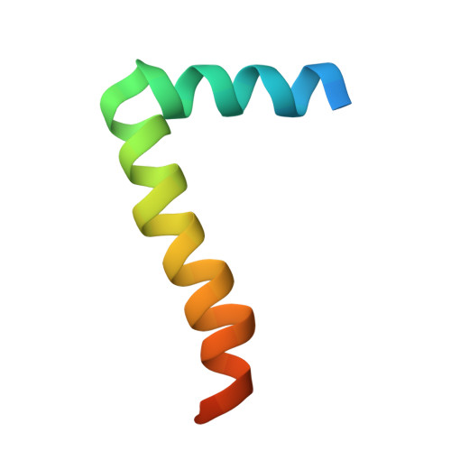
A solid-state NMR approach for simultaneous resonance assignment and three-dimensional structure determination of a membrane protein in lipid bilayers is described. The approach is based on the scattering, hence the descriptor "shotgun," of (15)N-labeled amino acids throughout the protein sequence (and the resulting NMR spectra). The samples are obtained by protein expression in bacteria grown on media in which one type of amino acid is labeled and the others are not. Shotgun NMR short-circuits the laborious and time-consuming process of obtaining complete sequential assignments prior to the calculation of a protein structure from the NMR data by taking advantage of the orientational information inherent to the spectra of aligned proteins. As a result, it is possible to simultaneously assign resonances and measure orientational restraints for structure determination. A total of five two-dimensional (1)H/(15)N PISEMA (polarization inversion spin exchange at the magic angle) spectra, from one uniformly and four selectively (15)N-labeled samples, were sufficient to determine the structure of the membrane-bound form of the 50-residue major pVIII coat protein of fd filamentous bacteriophage. Pisa (polarity index slat angle) wheels are an essential element in the process, which starts with the simultaneous assignment of resonances and the assembly of isolated polypeptide segments, and culminates in the complete three-dimensional structure of the protein with atomic resolution. The principles are also applicable to weakly aligned proteins studied by solution NMR spectroscopy. [The structure we determined for the membrane-bound form of the Fd bacteriophage pVIII coat protein has been deposited in the Protein Data Bank as PDB file 1MZT.]
Organizational Affiliation:
The Burnham Institute, La Jolla, California 92037, USA.