2ACQ

AN ANION BINDING SITE IN HUMAN ALDOSE REDUCTASE: MECHANISTIC IMPLICATIONS FOR THE BINDING OF CITRATE, CACODYLATE, AND GLUCOSE-6-PHOSPHATE


Experimental Data Snapshot

  • Method: X-RAY DIFFRACTION
  • Resolution: 1.76 Å
  • R-Value Work: 0.166 
  • R-Value Observed: 0.166 

wwPDB Validation   3D Report Full Report


This is version 1.4 of the entry. See complete history


Literature

An anion binding site in human aldose reductase: mechanistic implications for the binding of citrate, cacodylate, and glucose 6-phosphate.

Harrison, D.H.Bohren, K.M.Ringe, D.Petsko, G.A.Gabbay, K.H.

(1994) Biochemistry 33: 2011-2020

  • DOI: https://doi.org/10.1021/bi00174a006
  • Primary Citation of Related Structures:  
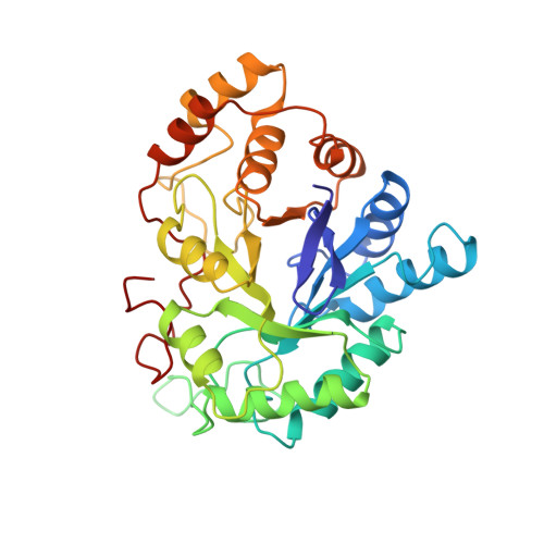
    2ACQ, 2ACR, 2ACS

  • PubMed Abstract: 

    Aldose reductase is a NADPH-dependent aldo-keto reductase involved in the pathogenesis of some diabetic and galactosemic complications. The published crystal structure of human aldose reductase [Wilson et al. (1992) Science 257, 81-84] contains a hitherto unexplained electron density positioned within the active site pocket facing the nicotinamide ring of the NADPH and other key active site residues (Tyr48, His110, and Cys298). In this paper we identify the electron density as citrate, which is present in the crystallization buffer (pH 5.0), and provide confirmatory evidence by both kinetic and crystallographic experiments. Citrate is an uncompetitive inhibitor in the forward reaction with respect to aldehyde (reduction of aldehyde), while it is a competitive inhibitor with respect to alcohol in the backward reaction (oxidation of alcohol), indicating that it interacts with the enzyme-NADP(+)-product complex. Citrate can be replaced in the crystalline enzyme complex by cacodylate or glucose 6-phosphate; the structure of each of these complexes shows the specific molecule bound in the active site. All of the structures have been determined to a nominal resolution of 1.76 A and refined to R-factors below 18%. While cacodylate can be bound within the active site under the crystallization conditions, it does not inhibit the wild-type enzyme in solution. Glucose 6-phosphate, however, is a substrate for aldose reductase. The similar location of the negative charges of citrate, cacodylate, and glucose 6-phosphate within the active site suggests an anion-binding site delineated by the C4N of nicotinamide, the OH of Tyr48, and the N epsilon of His110. The location of citrate binding in the active site leads to a plausible catalytic mechanism for aldose reductase.


  • Organizational Affiliation

    Department of Pediatrics, Baylor College of Medicine, Houston, Texas 77030.


Macromolecules
Find similar proteins by:  (by identity cutoff)  |  3D Structure
Entity ID: 1
MoleculeChains Sequence LengthOrganismDetailsImage
ALDOSE REDUCTASE315Homo sapiensMutation(s): 0 
EC: 1.1.1.21
UniProt & NIH Common Fund Data Resources
Find proteins for P15121 (Homo sapiens)
Explore P15121 
Go to UniProtKB:  P15121
PHAROS:  P15121
GTEx:  ENSG00000085662 
Entity Groups  
Sequence Clusters30% Identity50% Identity70% Identity90% Identity95% Identity100% Identity
UniProt GroupP15121
Sequence Annotations
Expand
  • Reference Sequence
Small Molecules
Ligands 2 Unique
IDChains Name / Formula / InChI Key2D Diagram3D Interactions
NAP
Query on NAP

Download Ideal Coordinates CCD File 
C [auth A]NADP NICOTINAMIDE-ADENINE-DINUCLEOTIDE PHOSPHATE
C21 H28 N7 O17 P3
XJLXINKUBYWONI-NNYOXOHSSA-N
G6P
Query on G6P

Download Ideal Coordinates CCD File 
B [auth A]6-O-phosphono-alpha-D-glucopyranose
C6 H13 O9 P
NBSCHQHZLSJFNQ-DVKNGEFBSA-N
Experimental Data & Validation

Experimental Data

  • Method: X-RAY DIFFRACTION
  • Resolution: 1.76 Å
  • R-Value Work: 0.166 
  • R-Value Observed: 0.166 
  • Space Group: P 21 21 21
Unit Cell:
Length ( Å )Angle ( ˚ )
a = 49.94α = 90
b = 67.17β = 90
c = 91.36γ = 90
Software Package:
Software NamePurpose
X-PLORmodel building
X-PLORrefinement
X-PLORphasing

Structure Validation

View Full Validation Report


Entry History 

Deposition Data

Revision History  (Full details and data files)

  • Version 1.0: 1994-07-31
    Type: Initial release
  • Version 1.1: 2008-03-03
    Changes: Version format compliance
  • Version 1.2: 2011-07-13
    Changes: Version format compliance
  • Version 1.3: 2020-07-29
    Type: Remediation
    Reason: Carbohydrate remediation
    Changes: Advisory, Data collection, Derived calculations, Other, Structure summary
  • Version 1.4: 2024-02-14
    Changes: Data collection, Database references, Structure summary