2AOA

Crystal structures of a high-affinity macrocyclic peptide mimetic in complex with the Grb2 SH2 domain


Experimental Data Snapshot

  • Method: X-RAY DIFFRACTION
  • Resolution: 1.99 Å
  • R-Value Free: 0.307 
  • R-Value Work: 0.246 
  • R-Value Observed: 0.249 

wwPDB Validation   3D Report Full Report


Ligand Structure Quality Assessment 


This is version 1.4 of the entry. See complete history


Literature

Crystal Structures of a High-affinity Macrocyclic Peptide Mimetic in Complex with the Grb2 SH2 Domain.

Phan, J.Shi, Z.D.Burke, T.R.Waugh, D.S.

(2005) J Mol Biol 353: 104-115

  • DOI: https://doi.org/10.1016/j.jmb.2005.08.037
  • Primary Citation of Related Structures:  
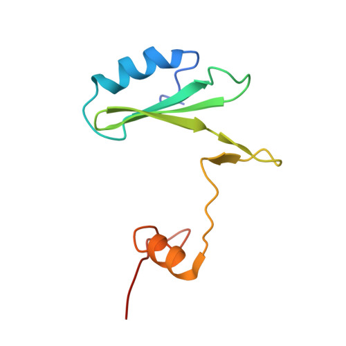
    2AOA, 2AOB

  • PubMed Abstract: 

    The high-affinity binding of the growth factor receptor-bound protein 2 (Grb2) SH2 domain to tyrosine-phosphorylated cytosolic domains of receptor tyrosine kinases (RTKs) is an attractive target for therapeutic intervention in many types of cancer. We report here two crystal forms of a complex between the Grb2 SH2 domain and a potent non-phosphorus-containing macrocyclic peptide mimetic that exhibits significant anti-proliferative effects against erbB-2-dependent breast cancers. This agent represents a "second generation" inhibitor with greatly improved binding affinity and bio-availability compared to its open-chain counterpart. The structures were determined at 2.0A and 1.8A with one and two domain-swapped dimers per asymmetric unit, respectively. The mode of binding and specific interactions between the protein and the inhibitor provide insight into the high potency of this class of macrocylic compounds and may aid in further optimization as part of the iterative rational drug design process.


  • Organizational Affiliation

    Macromolecular Crystallography Laboratory Center for Cancer Research, National Cancer Institute at Frederick, P.O. Box B, Frederick, MD 21702-1201, USA.


Macromolecules
Find similar proteins by:  (by identity cutoff)  |  3D Structure
Entity ID: 1
MoleculeChains Sequence LengthOrganismDetailsImage
Growth factor receptor-bound protein 2
A, B
99Homo sapiensMutation(s): 0 
Gene Names: GRB2ASH
UniProt & NIH Common Fund Data Resources
Find proteins for P62993 (Homo sapiens)
Explore P62993 
Go to UniProtKB:  P62993
PHAROS:  P62993
GTEx:  ENSG00000177885 
Entity Groups  
Sequence Clusters30% Identity50% Identity70% Identity90% Identity95% Identity100% Identity
UniProt GroupP62993
Sequence Annotations
Expand
  • Reference Sequence
Small Molecules
Ligands 2 Unique
IDChains Name / Formula / InChI Key2D Diagram3D Interactions
S1S
Query on S1S

Download Ideal Coordinates CCD File 
C [auth A],
D [auth A],
E [auth B]
2-(4-((9S,10S,14S,Z)-18-(2-AMINO-2-OXOETHYL)-9-(CARBOXYMETHYL)-14-(NAPHTHALEN-1-YLMETHYL)-8,17,20-TRIOXO-7,16,19-TRIAZASPIRO[5.14]ICOS-11-EN-10-YL)PHENYL)MALONIC ACID
C41 H46 N4 O10
FYIJOFSDJMOTGX-HGOOHLDYSA-N
P33
Query on P33

Download Ideal Coordinates CCD File 
F [auth B]3,6,9,12,15,18-HEXAOXAICOSANE-1,20-DIOL
C14 H30 O8
XPJRQAIZZQMSCM-UHFFFAOYSA-N
Experimental Data & Validation

Experimental Data

  • Method: X-RAY DIFFRACTION
  • Resolution: 1.99 Å
  • R-Value Free: 0.307 
  • R-Value Work: 0.246 
  • R-Value Observed: 0.249 
  • Space Group: F 2 2 2
Unit Cell:
Length ( Å )Angle ( ˚ )
a = 88.173α = 90
b = 102.659β = 90
c = 107.24γ = 90
Software Package:
Software NamePurpose
REFMACrefinement
MAR345data collection
SCALEPACKdata scaling
AMoREphasing

Structure Validation

View Full Validation Report


Ligand Structure Quality Assessment 


Entry History 

Deposition Data

Revision History  (Full details and data files)

  • Version 1.0: 2005-10-04
    Type: Initial release
  • Version 1.1: 2008-04-30
    Changes: Version format compliance
  • Version 1.2: 2011-07-13
    Changes: Derived calculations, Version format compliance
  • Version 1.3: 2017-10-11
    Changes: Refinement description
  • Version 1.4: 2024-02-14
    Changes: Data collection, Database references, Derived calculations, Structure summary