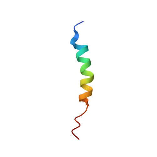
Contrast-matched small-angle X-ray scattering from a heavy-atom-labeled protein in structure determination: application to a lead-substituted calmodulin-peptide complex.
Grishaev, A., Anthis, N.J., Clore, G.M.(2012) J Am Chem Soc 134: 14686-14689
- PubMed: 22908850
- DOI: https://doi.org/10.1021/ja306359z
- Primary Citation of Related Structures:
2LV6 - PubMed Abstract:
The information content in 1-D solution X-ray scattering profiles is generally restricted to low-resolution shape and size information that, on its own, cannot lead to unique 3-D structures of biological macromolecules comparable to all-atom models derived from X-ray crystallography or NMR spectroscopy. Here we show that contrast-matched X-ray scattering data collected on a protein incorporating specific heavy-atom labels in 65% aqueous sucrose buffer can dramatically enhance the power of conventional small- and wide-angle X-ray scattering (SAXS/WAXS) measurements. Under contrast-matching conditions the protein is effectively invisible and the main contribution to the X-ray scattering intensity arises from the heavy atoms, allowing direct extraction of pairwise distances between them. In combination with conventional aqueous SAXS/WAXS data, supplemented by NMR-derived residual dipolar couplings (RDCs) measured in a weakly aligning medium, we show that it is possible to position protein domains relative to one another within a precision of 1 Å. We demonstrate this approach with respect to the determination of domain positions in a complex between calmodulin, in which the four Ca(2+) ions have been substituted by Pb(2+), and a target peptide. The uniqueness of the resulting solution is established by an exhaustive search over all models compatible with the experimental data, and could not have been achieved using aqueous SAXS and RDC data alone. Moreover, we show that the correct structural solution can be recovered using only contrast-matched SAXS and aqueous SAXS/WAXS data.
Organizational Affiliation:
Laboratories of Chemical Physics, National Institute of Diabetes and Digestive and Kidney Diseases, National Institutes of Health, Bethesda, Maryland 20892-0520, USA.