2Q9E

Structure of spin-labeled T4 lysozyme mutant S44R1


Experimental Data Snapshot

  • Method: X-RAY DIFFRACTION
  • Resolution: 2.10 Å
  • R-Value Free: 0.250 
  • R-Value Work: 0.202 

  • Method: EPR

wwPDB Validation   3D Report Full Report


Ligand Structure Quality Assessment 


This is version 1.6 of the entry. See complete history


Literature

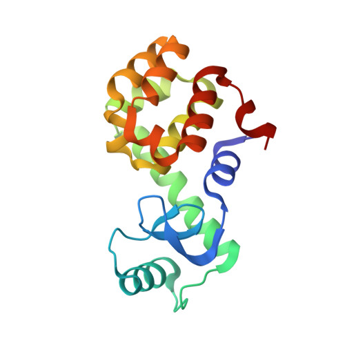
Structural determinants of nitroxide motion in spin-labeled proteins: Solvent-exposed sites in helix B of T4 lysozyme.

Guo, Z.Cascio, D.Hideg, K.Hubbell, W.L.

(2008) Protein Sci 17: 228-239

  • DOI: https://doi.org/10.1110/ps.073174008
  • Primary Citation of Related Structures:  
    2Q9D, 2Q9E

  • PubMed Abstract: 

    Site-directed spin labeling provides a means for exploring structure and dynamics in proteins. To interpret the complex EPR spectra that often arise, it is necessary to characterize the rotamers of the spin-labeled side chain and the interactions they make with the local environment in proteins of known structure. For this purpose, crystal structures have been determined for T4 lysozyme bearing a nitroxide side chain (R1) at the solvent-exposed helical sites 41 and 44 in the B helix. These sites are of particular interest in that the corresponding EPR spectra reveal two dynamic states of R1, one of which is relatively immobilized suggesting interactions of the nitroxide with the environment. The crystal structures together with the effect of mutagenesis of nearest neighbors on the motion of R1 suggest intrahelical interactions of 41R1 with the i + 4 residue and of 44R1 with the i + 1 residue. Such interactions appear to be specific to particular rotamers of the R1 side chain.


  • Organizational Affiliation

    Jules Stein Eye Institute and Department of Chemistry and Biochemistry, University of California, Los Angeles, California 90095-7008, USA.


Macromolecules
Find similar proteins by:  (by identity cutoff)  |  3D Structure
Entity ID: 1
MoleculeChains Sequence LengthOrganismDetailsImage
Lysozyme
A, B, C
164Tequatrovirus T4Mutation(s): 4 
Gene Names: E
EC: 3.2.1.17
UniProt
Find proteins for P00720 (Enterobacteria phage T4)
Explore P00720 
Go to UniProtKB:  P00720
Entity Groups  
Sequence Clusters30% Identity50% Identity70% Identity90% Identity95% Identity100% Identity
UniProt GroupP00720
Sequence Annotations
Expand
  • Reference Sequence
Small Molecules
Experimental Data & Validation

Experimental Data

  • Method: X-RAY DIFFRACTION
  • Resolution: 2.10 Å
  • R-Value Free: 0.250 
  • R-Value Work: 0.202 
  • Space Group: P 21 21 21
Unit Cell:
Length ( Å )Angle ( ˚ )
a = 57.989α = 90
b = 67.14β = 90
c = 165.919γ = 90
Software Package:
Software NamePurpose
DENZOdata reduction
SCALEPACKdata scaling
CNSrefinement
PDB_EXTRACTdata extraction
ADSCdata collection
MOLREPphasing

Structure Validation

View Full Validation Report


Ligand Structure Quality Assessment 


Entry History 

Deposition Data

Revision History  (Full details and data files)

  • Version 1.0: 2007-06-26
    Type: Initial release
  • Version 1.1: 2008-02-05
    Changes: Version format compliance
  • Version 1.2: 2011-07-13
    Changes: Version format compliance
  • Version 1.3: 2011-07-27
    Changes: Non-polymer description
  • Version 1.4: 2017-10-18
    Changes: Advisory, Refinement description
  • Version 1.5: 2021-10-20
    Changes: Database references, Derived calculations
  • Version 1.6: 2023-08-30
    Changes: Data collection, Refinement description