2QUZ

Crystal Structure of the activating H-RasK117R mutant in Costello Syndrome, bound to Mg-GDP


Experimental Data Snapshot

  • Method: X-RAY DIFFRACTION
  • Resolution: 1.49 Å
  • R-Value Free: 0.222 
  • R-Value Work: 0.203 
  • R-Value Observed: 0.204 

wwPDB Validation   3D Report Full Report


Ligand Structure Quality Assessment 


This is version 1.3 of the entry. See complete history


Literature

Mutation analysis in Costello syndrome: functional and structural characterization of the HRAS p.Lys117Arg mutation.

Denayer, E.Parret, A.Chmara, M.Schubbert, S.Vogels, A.Devriendt, K.Frijns, J.P.Rybin, V.de Ravel, T.J.Shannon, K.Cools, J.Scheffzek, K.Legius, E.

(2008) Hum Mutat 29: 232-239

  • DOI: https://doi.org/10.1002/humu.20616
  • Primary Citation of Related Structures:  
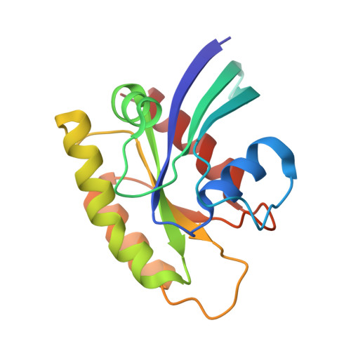
    2QUZ

  • PubMed Abstract: 

    Costello syndrome is a mental retardation syndrome characterized by high birth weight, postnatal growth retardation, coarse face, loose skin, cardiovascular problems, and tumor predisposition. De novo heterozygous missense mutations in HRAS codon 12 and 13 disturbing the intrinsic GTP hydrolysis cause Costello syndrome. We report a patient with typical Costello syndrome and a novel heterozygous missense mutation in codon 117 (c.350A>G, p.Lys117Arg) of the HRAS gene, resulting in constitutive activation of the RAS/MAPK pathway similar to the typical p.Gly12Ser and p.Gly12Ala mutations. Recombinant HRAS p.Lys117Arg demonstrates normal intrinsic GTP hydrolysis and responsiveness to GTPase-activating proteins, but the nucleotide dissociation rate is increased 80-fold. Consistent with the biochemical data, the crystal structure of the p.Lys117Arg mutant indicates an altered interaction pattern of the side chain that is associated with unfavorable nucleotide binding properties. Together, these data show that a RAS mutation that only perturbs guanine nucleotide binding has similar functional consequences as mutations that impair GTP hydrolysis and causes human disease.


  • Organizational Affiliation

    Department of Human Genetics, Catholic University of Leuven, Leuven, Belgium.


Macromolecules
Find similar proteins by:  (by identity cutoff)  |  3D Structure
Entity ID: 1
MoleculeChains Sequence LengthOrganismDetailsImage
GTPase HRas166Homo sapiensMutation(s): 1 
Gene Names: HRASHRAS1
EC: 3.6.5.2
UniProt & NIH Common Fund Data Resources
Find proteins for P01112 (Homo sapiens)
Explore P01112 
Go to UniProtKB:  P01112
PHAROS:  P01112
GTEx:  ENSG00000174775 
Entity Groups  
Sequence Clusters30% Identity50% Identity70% Identity90% Identity95% Identity100% Identity
UniProt GroupP01112
Sequence Annotations
Expand
  • Reference Sequence
Small Molecules
Ligands 2 Unique
IDChains Name / Formula / InChI Key2D Diagram3D Interactions
GDP
Query on GDP

Download Ideal Coordinates CCD File 
C [auth A]GUANOSINE-5'-DIPHOSPHATE
C10 H15 N5 O11 P2
QGWNDRXFNXRZMB-UUOKFMHZSA-N
MG
Query on MG

Download Ideal Coordinates CCD File 
B [auth A]MAGNESIUM ION
Mg
JLVVSXFLKOJNIY-UHFFFAOYSA-N
Experimental Data & Validation

Experimental Data

  • Method: X-RAY DIFFRACTION
  • Resolution: 1.49 Å
  • R-Value Free: 0.222 
  • R-Value Work: 0.203 
  • R-Value Observed: 0.204 
  • Space Group: H 3 2
Unit Cell:
Length ( Å )Angle ( ˚ )
a = 92.1α = 90
b = 92.1β = 90
c = 116.3γ = 120
Software Package:
Software NamePurpose
XSCALEdata scaling
PHASERphasing
REFMACrefinement
PDB_EXTRACTdata extraction
ADSCdata collection
XDSdata reduction

Structure Validation

View Full Validation Report


Ligand Structure Quality Assessment 


Entry History 

Deposition Data

Revision History  (Full details and data files)

  • Version 1.0: 2007-12-11
    Type: Initial release
  • Version 1.1: 2011-07-13
    Changes: Version format compliance
  • Version 1.2: 2021-10-20
    Changes: Database references, Derived calculations
  • Version 1.3: 2023-08-30
    Changes: Data collection, Refinement description