3NOB

Structure of K11-linked di-ubiquitin


Experimental Data Snapshot

  • Method: X-RAY DIFFRACTION
  • Resolution: 2.19 Å
  • R-Value Free: 0.281 
  • R-Value Work: 0.245 
  • R-Value Observed: 0.247 

wwPDB Validation   3D Report Full Report


This is version 1.3 of the entry. See complete history


Literature

K11-Linked Polyubiquitination in Cell Cycle Control Revealed by a K11 Linkage-Specific Antibody.

Matsumoto, M.L.Wickliffe, K.E.Dong, K.C.Yu, C.Bosanac, I.Bustos, D.Phu, L.Kirkpatrick, D.S.Hymowitz, S.G.Rape, M.Kelley, R.F.Dixit, V.M.

(2010) Mol Cell 39: 477-484

  • DOI: https://doi.org/10.1016/j.molcel.2010.07.001
  • Primary Citation of Related Structures:  
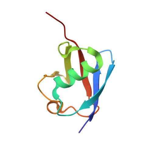
    3NOB

  • PubMed Abstract: 

    Polyubiquitination is a posttranslational modification where ubiquitin chains containing isopeptide bonds linking one of seven ubiquitin lysines with the C terminus of an adjoining ubiquitin are covalently attached to proteins. While functions of K48- and K63-linked polyubiquitin are understood, the role(s) of noncanonical K11-linked chains is less clear. A crystal structure of K11-linked diubiquitin demonstrates a distinct conformation from K48- or K63-linked diubiquitin. We engineered a K11 linkage-specific antibody and use it to demonstrate that K11 chains are highly upregulated in mitotic human cells precisely when substrates of the ubiquitin ligase anaphase-promoting complex (APC/C) are degraded. These chains increased with proteasomal inhibition, suggesting they act as degradation signals in vivo. Inhibition of the APC/C strongly impeded the formation of K11-linked chains, suggesting that a single ubiquitin ligase is the major source of mitotic K11-linked chains. Our results underscore the importance of K11-linked ubiquitin chains as critical regulators of mitotic protein degradation.


  • Organizational Affiliation

    Department of Antibody Engineering, Genentech, Inc., South San Francisco, CA 94080, USA.


Macromolecules
Find similar proteins by:  (by identity cutoff)  |  3D Structure
Entity ID: 1
MoleculeChains Sequence LengthOrganismDetailsImage
Ubiquitin
A, B, C, D, E
A, B, C, D, E, F, G, H
78Homo sapiensMutation(s): 0 
Gene Names: RPS27AUBA80UBCEP1UBA52UBBUBC
UniProt & NIH Common Fund Data Resources
Find proteins for P62979 (Homo sapiens)
Explore P62979 
Go to UniProtKB:  P62979
PHAROS:  P62979
GTEx:  ENSG00000143947 
Entity Groups  
Sequence Clusters30% Identity50% Identity70% Identity90% Identity95% Identity100% Identity
UniProt GroupP62979
Sequence Annotations
Expand
  • Reference Sequence
Experimental Data & Validation

Experimental Data

  • Method: X-RAY DIFFRACTION
  • Resolution: 2.19 Å
  • R-Value Free: 0.281 
  • R-Value Work: 0.245 
  • R-Value Observed: 0.247 
  • Space Group: C 1 2 1
Unit Cell:
Length ( Å )Angle ( ˚ )
a = 155.597α = 90
b = 56.059β = 95.21
c = 65.772γ = 90
Software Package:
Software NamePurpose
BOSdata collection
PHASERphasing
PHENIXrefinement
HKL-3000data reduction
HKL-3000data scaling

Structure Validation

View Full Validation Report


Entry History 

Deposition Data

Revision History  (Full details and data files)

  • Version 1.0: 2010-08-11
    Type: Initial release
  • Version 1.1: 2011-07-13
    Changes: Version format compliance
  • Version 1.2: 2014-04-02
    Changes: Structure summary
  • Version 1.3: 2023-09-06
    Changes: Data collection, Database references, Derived calculations, Refinement description