


Crystal Structure of Ribosomal Protein L1 from the Bacterium Aquifex Aeolicus
Nikonova, E.Yu., Tishchenko, S.V., Gabdulkhakov, A.G., Shklyaeva, A.A., Garber, M.B., Nikonov, S.V., Nevskaya, N.A.(2011) Crystallogr Rep 56: 603-607
Experimental Data Snapshot
wwPDB Validation 3D Report Full Report

(2011) Crystallogr Rep 56: 603-607
Entity ID: 1 | |||||
|---|---|---|---|---|---|
| Molecule | Chains | Sequence Length | Organism | Details | Image |
| 50S ribosomal protein L1 | 242 | Aquifex aeolicus | Mutation(s): 0 Gene Names: aq_1935, rplA | 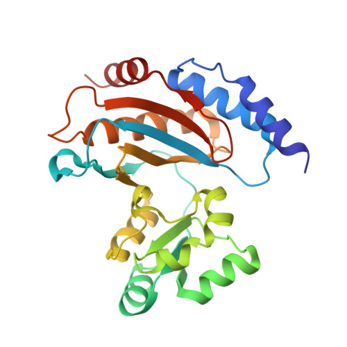 | |
UniProt | |||||
Find proteins for O67759 (Aquifex aeolicus (strain VF5)) Explore O67759 Go to UniProtKB: O67759 | |||||
Entity Groups | |||||
| Sequence Clusters | 30% Identity50% Identity70% Identity90% Identity95% Identity100% Identity | ||||
| UniProt Group | O67759 | ||||
Sequence AnnotationsExpand | |||||
| |||||
| Ligands 3 Unique | |||||
|---|---|---|---|---|---|
| ID | Chains | Name / Formula / InChI Key | 2D Diagram | 3D Interactions | |
| TRS Query on TRS | D [auth A] | 2-AMINO-2-HYDROXYMETHYL-PROPANE-1,3-DIOL C4 H12 N O3 LENZDBCJOHFCAS-UHFFFAOYSA-O |  | ||
| SO4 Query on SO4 | B [auth A], C [auth A] | SULFATE ION O4 S QAOWNCQODCNURD-UHFFFAOYSA-L |  | ||
| ACY Query on ACY | E [auth A] | ACETIC ACID C2 H4 O2 QTBSBXVTEAMEQO-UHFFFAOYSA-N |  | ||
| Length ( Å ) | Angle ( ˚ ) |
|---|---|
| a = 107.49 | α = 90 |
| b = 37.52 | β = 110.32 |
| c = 58.97 | γ = 90 |
| Software Name | Purpose |
|---|---|
| MAR345dtb | data collection |
| PHASER | phasing |
| PHENIX | refinement |
| XDS | data reduction |
| XDS | data scaling |
RCSB PDB (citation) is hosted by
RCSB PDB is a member of the

