4FJV

Crystal Structure of Human Otubain2 and Ubiquitin Complex


Experimental Data Snapshot

  • Method: X-RAY DIFFRACTION
  • Resolution: 2.05 Å
  • R-Value Free: 0.267 
  • R-Value Work: 0.206 
  • R-Value Observed: 0.209 

wwPDB Validation   3D Report Full Report


This is version 1.2 of the entry. See complete history


Literature

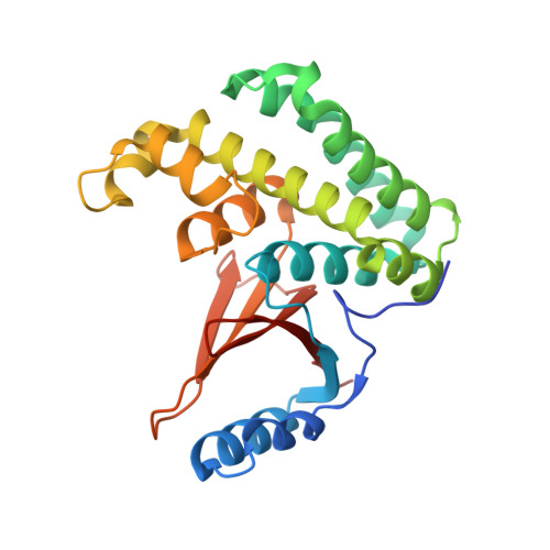
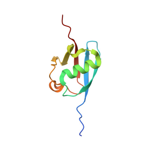
The human otubain2-ubiquitin structure provides insights into the cleavage specificity of poly-ubiquitin-linkages.

Altun, M.Walter, T.S.Kramer, H.B.Herr, P.Iphofer, A.Bostrom, J.David, Y.Komsany, A.Ternette, N.Navon, A.Stuart, D.I.Ren, J.Kessler, B.M.

(2015) PLoS One 10: e0115344-e0115344

  • DOI: https://doi.org/10.1371/journal.pone.0115344
  • Primary Citation of Related Structures:  
    4FJV

  • PubMed Abstract: 

    Ovarian tumor domain containing proteases cleave ubiquitin (Ub) and ubiquitin-like polypeptides from proteins. Here we report the crystal structure of human otubain 2 (OTUB2) in complex with a ubiquitin-based covalent inhibitor, Ub-Br2. The ubiquitin binding mode is oriented differently to how viral otubains (vOTUs) bind ubiquitin/ISG15, and more similar to yeast and mammalian OTUs. In contrast to OTUB1 which has exclusive specificity towards Lys48 poly-ubiquitin chains, OTUB2 cleaves different poly-Ub linked chains. N-terminal tail swapping experiments between OTUB1 and OTUB2 revealed how the N-terminal structural motifs in OTUB1 contribute to modulating enzyme activity and Ub-chain selectivity, a trait not observed in OTUB2, supporting the notion that OTUB2 may affect a different spectrum of substrates in Ub-dependent pathways.


  • Organizational Affiliation

    Target Discovery Institute, Nuffield Department of Medicine, Roosevelt Drive, University of Oxford, Oxford, OX3 7FZ, United Kingdom; Science for Life Laboratory, Division of Translational Medicine and Chemical Biology, Department of Medical Biochemistry and Biophysics, Karolinska Institutet, SE-171 21 Stockholm, Sweden.


Macromolecules
Find similar proteins by:  (by identity cutoff)  |  3D Structure
Entity ID: 1
MoleculeChains Sequence LengthOrganismDetailsImage
Ubiquitin thioesterase OTUB2
A, C
237Homo sapiensMutation(s): 0 
Gene Names: OTUB2C14orf137OTB2OTU2
EC: 3.4.19.12
UniProt & NIH Common Fund Data Resources
Find proteins for Q96DC9 (Homo sapiens)
Explore Q96DC9 
Go to UniProtKB:  Q96DC9
PHAROS:  Q96DC9
GTEx:  ENSG00000089723 
Entity Groups  
Sequence Clusters30% Identity50% Identity70% Identity90% Identity95% Identity100% Identity
UniProt GroupQ96DC9
Sequence Annotations
Expand
  • Reference Sequence
Find similar proteins by:  (by identity cutoff)  |  3D Structure
Entity ID: 2
MoleculeChains Sequence LengthOrganismDetailsImage
Polyubiquitin-C
B, D
86Homo sapiensMutation(s): 0 
Gene Names: UBC
UniProt & NIH Common Fund Data Resources
Find proteins for P0CG48 (Homo sapiens)
Explore P0CG48 
Go to UniProtKB:  P0CG48
PHAROS:  P0CG48
GTEx:  ENSG00000150991 
Entity Groups  
Sequence Clusters30% Identity50% Identity70% Identity90% Identity95% Identity100% Identity
UniProt GroupP0CG48
Sequence Annotations
Expand
  • Reference Sequence
Experimental Data & Validation

Experimental Data

  • Method: X-RAY DIFFRACTION
  • Resolution: 2.05 Å
  • R-Value Free: 0.267 
  • R-Value Work: 0.206 
  • R-Value Observed: 0.209 
  • Space Group: P 21 21 21
Unit Cell:
Length ( Å )Angle ( ˚ )
a = 54.063α = 90
b = 76.825β = 90
c = 198.847γ = 90
Software Package:
Software NamePurpose
MOLREPphasing
PHENIXrefinement
HKL-2000data reduction
SCALEPACKdata scaling

Structure Validation

View Full Validation Report


Entry History 

Deposition Data

Revision History  (Full details and data files)

  • Version 1.0: 2013-06-12
    Type: Initial release
  • Version 1.1: 2015-02-25
    Changes: Database references
  • Version 1.2: 2023-09-13
    Changes: Data collection, Database references, Derived calculations, Refinement description