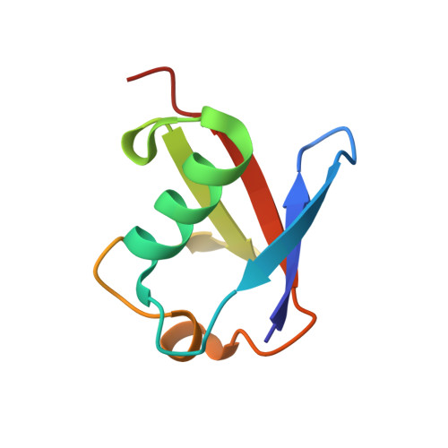
Assembly and structure of Lys33-linked polyubiquitin reveals distinct conformations.
Kristariyanto, Y.A., Choi, S.Y., Rehman, S.A., Ritorto, M.S., Campbell, D.G., Morrice, N.A., Toth, R., Kulathu, Y.(2015) Biochem J 467: 345-352
- PubMed: 25723849
- DOI: https://doi.org/10.1042/BJ20141502
- Primary Citation of Related Structures:
4XYZ, 4Y1H - PubMed Abstract:
Ubiquitylation regulates a multitude of biological processes and this versatility stems from the ability of ubiquitin (Ub) to form topologically different polymers of eight different linkage types. Whereas some linkages have been studied in detail, other linkage types including Lys33-linked polyUb are poorly understood. In the present study, we identify an enzymatic system for the large-scale assembly of Lys33 chains by combining the HECT (homologous to the E6-AP C-terminus) E3 ligase AREL1 (apoptosis-resistant E3 Ub protein ligase 1) with linkage selective deubiquitinases (DUBs). Moreover, this first characterization of the chain selectivity of AREL1 indicates its preference for assembling Lys33- and Lys11-linked Ub chains. Intriguingly, the crystal structure of Lys33-linked diUb reveals that it adopts a compact conformation very similar to that observed for Lys11-linked diUb. In contrast, crystallographic analysis of Lys33-linked triUb reveals a more extended conformation. These two distinct conformational states of Lys33-linked polyUb may be selectively recognized by Ub-binding domains (UBD) and enzymes of the Ub system. Importantly, our work provides a method to assemble Lys33-linked polyUb that will allow further characterization of this atypical chain type.
Organizational Affiliation:
*MRC Protein Phosphorylation and Ubiquitylation Unit, College of Life Sciences, University of Dundee, Dow Street, Dundee DD1 5EH, U.K.