5ID6

Structure of Cpf1/RNA Complex


Experimental Data Snapshot

  • Method: X-RAY DIFFRACTION
  • Resolution: 2.38 Å
  • R-Value Free: 0.261 
  • R-Value Work: 0.202 
  • R-Value Observed: 0.205 

wwPDB Validation   3D Report Full Report


This is version 1.2 of the entry. See complete history


Literature

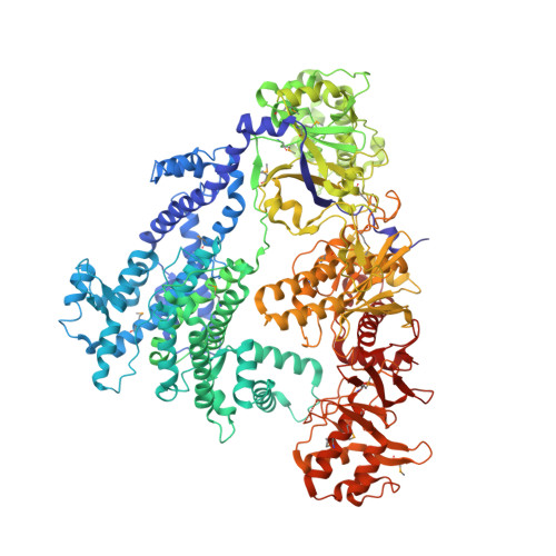
The crystal structure of Cpf1 in complex with CRISPR RNA

Dong, D.Ren, K.Qiu, X.Zheng, J.Guo, M.Guan, X.Liu, H.Li, N.Zhang, B.Yang, D.Ma, C.Wang, S.Wu, D.Ma, Y.Fan, S.Wang, J.Gao, N.Huang, Z.

(2016) Nature 532: 522-526

  • DOI: https://doi.org/10.1038/nature17944
  • Primary Citation of Related Structures:  
    5ID6

  • PubMed Abstract: 

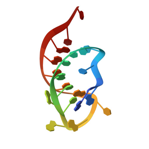
    The CRISPR-Cas systems, as exemplified by CRISPR-Cas9, are RNA-guided adaptive immune systems used by bacteria and archaea to defend against viral infection. The CRISPR-Cpf1 system, a new class 2 CRISPR-Cas system, mediates robust DNA interference in human cells. Although functionally conserved, Cpf1 and Cas9 differ in many aspects including their guide RNAs and substrate specificity. Here we report the 2.38 Å crystal structure of the CRISPR RNA (crRNA)-bound Lachnospiraceae bacterium ND2006 Cpf1 (LbCpf1). LbCpf1 has a triangle-shaped architecture with a large positively charged channel at the centre. Recognized by the oligonucleotide-binding domain of LbCpf1, the crRNA adopts a highly distorted conformation stabilized by extensive intramolecular interactions and the (Mg(H2O)6)(2+) ion. The oligonucleotide-binding domain also harbours a looped-out helical domain that is important for LbCpf1 substrate binding. Binding of crRNA or crRNA lacking the guide sequence induces marked conformational changes but no oligomerization of LbCpf1. Our study reveals the crRNA recognition mechanism and provides insight into crRNA-guided substrate binding of LbCpf1, establishing a framework for engineering LbCpf1 to improve its efficiency and specificity for genome editing.


  • Organizational Affiliation

    School of Life Science and Technology, Harbin Institute of Technology, Harbin 150080, China.


Macromolecules

Find similar proteins by:  (by identity cutoff)  |  3D Structure
Entity ID: 1
MoleculeChains Sequence LengthOrganismDetailsImage
Cpf11,228Lachnospiraceae bacterium ND2006Mutation(s): 0 
UniProt
Find proteins for A0A182DWE3 (Lachnospiraceae bacterium ND2006)
Explore A0A182DWE3 
Go to UniProtKB:  A0A182DWE3
Entity Groups  
Sequence Clusters30% Identity50% Identity70% Identity90% Identity95% Identity100% Identity
UniProt GroupA0A182DWE3
Sequence Annotations
Expand
  • Reference Sequence

Find similar nucleic acids by:  Sequence   |   3D Structure  

Entity ID: 2
MoleculeChains LengthOrganismImage
RNA (5'-R(P*AP*AP*UP*UP*UP*CP*UP*AP*CP*UP*AP*AP*GP*UP*GP*UP*AP*GP*AP*UP*C)-3')B [auth G]21synthetic construct
Sequence Annotations
Expand
  • Reference Sequence
Small Molecules
Modified Residues  1 Unique
IDChains TypeFormula2D DiagramParent
MSE
Query on MSE
A
L-PEPTIDE LINKINGC5 H11 N O2 SeMET
Experimental Data & Validation

Experimental Data

  • Method: X-RAY DIFFRACTION
  • Resolution: 2.38 Å
  • R-Value Free: 0.261 
  • R-Value Work: 0.202 
  • R-Value Observed: 0.205 
  • Space Group: C 1 2 1
Unit Cell:
Length ( Å )Angle ( ˚ )
a = 168.352α = 90
b = 83.011β = 106.72
c = 124.086γ = 90
Software Package:
Software NamePurpose
PHENIXrefinement
HKL-2000data scaling
PDB_EXTRACTdata extraction
Cootmodel building

Structure Validation

View Full Validation Report


Entry History 

Deposition Data

Revision History  (Full details and data files)

  • Version 1.0: 2016-04-27
    Type: Initial release
  • Version 1.1: 2016-05-04
    Changes: Database references
  • Version 1.2: 2016-05-11
    Changes: Database references