5JRQ

BRAFV600E Kinase Domain In Complex with Chemically Linked Vemurafenib Inhibitor VEM-6-VEM


Experimental Data Snapshot

  • Method: X-RAY DIFFRACTION
  • Resolution: 2.29 Å
  • R-Value Free: 0.254 
  • R-Value Work: 0.195 
  • R-Value Observed: 0.200 

wwPDB Validation   3D Report Full Report


Ligand Structure Quality Assessment 


This is version 2.1 of the entry. See complete history


Literature

Chemically Linked Vemurafenib Inhibitors Promote an Inactive BRAF(V600E) Conformation.

Grasso, M.Estrada, M.A.Ventocilla, C.Samanta, M.Maksimoska, J.Villanueva, J.Winkler, J.D.Marmorstein, R.

(2016) ACS Chem Biol 11: 2876-2888

  • DOI: https://doi.org/10.1021/acschembio.6b00529
  • Primary Citation of Related Structures:  
    5JRQ, 5JSM, 5JT2

  • PubMed Abstract: 

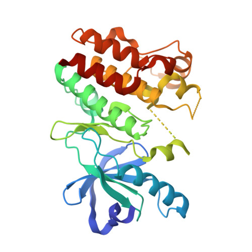
    The BRAF kinase, within the mitogen activated protein kinase (MAPK) signaling pathway, harbors activating mutations in about half of melanomas and to a significant extent in many other cancers. A single valine to glutamic acid substitution at residue 600 (BRAF V600E ) accounts for about 90% of these activating mutations. While BRAF V600E -selective small molecule inhibitors, such as debrafenib and vemurafenib, have shown therapeutic benefit, almost all patients develop resistance. Resistance often arises through reactivation of the MAPK pathway, typically through mutation of upstream RAS, downstream MEK, or splicing variants. RAF kinases signal as homo- and heterodimers, and another complication associated with small molecule BRAF V600E inhibition is drug-induced allosteric activation of a wild-type RAF subunit (BRAF or CRAF) of the kinase dimer, a process called "transactivation" or "paradoxical activation." Here, we used BRAF V600E and vemurafenib as a model system to develop chemically linked kinase inhibitors to lock RAF dimers in an inactive conformation that cannot undergo transactivation. This structure-based design effort resulted in the development of Vem-BisAmide-2, a compound containing two vemurafenib molecules connected by a bis amide linker. We show that Vem-BisAmide-2 has comparable inhibitory potency as vemurafenib to BRAF V600E both in vitro and in cells but promotes an inactive dimeric BRAF V600E conformation unable to undergo transactivation. The crystal structure of a BRAF V600E /Vem-BisAmide-2 complex and associated biochemical studies reveal the molecular basis for how Vem-BisAmide-2 mediates selectivity for an inactive over an active dimeric BRAF V600E conformation. These studies have implications for targeting BRAF V600E /RAF heterodimers and other kinase dimers for therapy.


  • Organizational Affiliation

    Department of Biochemistry and Biophysics and the Abramson Family Cancer Research Institute, Perelman School of Medicine, University of Pennsylvania , 421 Curie Blvd., Philadelphia, Pennsylvania 19104, United States.


Macromolecules
Find similar proteins by:  (by identity cutoff)  |  3D Structure
Entity ID: 1
MoleculeChains Sequence LengthOrganismDetailsImage
Serine/threonine-protein kinase B-raf
A, B
280Homo sapiensMutation(s): 17 
Gene Names: BRAFBRAF1RAFB1
EC: 2.7.11.1
UniProt & NIH Common Fund Data Resources
Find proteins for P15056 (Homo sapiens)
Explore P15056 
Go to UniProtKB:  P15056
PHAROS:  P15056
GTEx:  ENSG00000157764 
Entity Groups  
Sequence Clusters30% Identity50% Identity70% Identity90% Identity95% Identity100% Identity
UniProt GroupP15056
Sequence Annotations
Expand
  • Reference Sequence
Small Molecules
Ligands 4 Unique
IDChains Name / Formula / InChI Key2D Diagram3D Interactions
6N9
Query on 6N9

Download Ideal Coordinates CCD File 
C [auth A],
G [auth B]
N-{2,4-difluoro-3-[5-(4-methoxyphenyl)-1H-pyrrolo[2,3-b]pyridine-3-carbonyl]phenyl}propane-1-sulfonamide
C24 H21 F2 N3 O4 S
WEEVBQHRUFJEAI-UHFFFAOYSA-N
GOL
Query on GOL

Download Ideal Coordinates CCD File 
E [auth A]GLYCEROL
C3 H8 O3
PEDCQBHIVMGVHV-UHFFFAOYSA-N
DMS
Query on DMS

Download Ideal Coordinates CCD File 
D [auth A],
H [auth B]
DIMETHYL SULFOXIDE
C2 H6 O S
IAZDPXIOMUYVGZ-UHFFFAOYSA-N
TMA
Query on TMA

Download Ideal Coordinates CCD File 
F [auth A],
I [auth B]
TETRAMETHYLAMMONIUM ION
C4 H12 N
QEMXHQIAXOOASZ-UHFFFAOYSA-N
Experimental Data & Validation

Experimental Data

  • Method: X-RAY DIFFRACTION
  • Resolution: 2.29 Å
  • R-Value Free: 0.254 
  • R-Value Work: 0.195 
  • R-Value Observed: 0.200 
  • Space Group: P 1 21 1
Unit Cell:
Length ( Å )Angle ( ˚ )
a = 56.836α = 90
b = 67.753β = 90.32
c = 67.881γ = 90
Software Package:
Software NamePurpose
PHENIXrefinement
HKL-3000data reduction
HKL-3000data scaling
PHASERphasing
PDB_EXTRACTdata extraction
Cootmodel building

Structure Validation

View Full Validation Report


Ligand Structure Quality Assessment 


Entry History & Funding Information

Deposition Data


Funding OrganizationLocationGrant Number
National Institutes of Health/National Cancer Institute (NIH/NCI)United StatesCA114046

Revision History  (Full details and data files)

  • Version 1.0: 2016-09-14
    Type: Initial release
  • Version 1.1: 2016-09-21
    Changes: Database references
  • Version 1.2: 2016-11-02
    Changes: Database references
  • Version 1.3: 2017-09-27
    Changes: Author supporting evidence, Derived calculations
  • Version 1.4: 2019-12-04
    Changes: Author supporting evidence
  • Version 2.0: 2022-11-02
    Changes: Atomic model, Database references, Derived calculations, Non-polymer description, Structure summary
  • Version 2.1: 2023-10-18
    Changes: Data collection, Refinement description