Structural and functional properties of Grb2 SH2 dimer in CD28 binding.
Hosoe, Y., Numoto, N., Inaba, S., Ogawa, S., Morii, H., Abe, R., Ito, N., Oda, M.(2019) Biophys Physicobiol 16: 80-88
- PubMed: 30923665
- DOI: https://doi.org/10.2142/biophysico.16.0_80
- Primary Citation of Related Structures:
6ICG, 6ICH - PubMed Abstract:
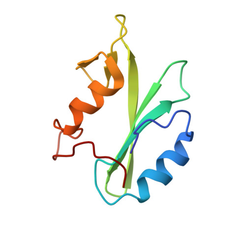
Growth factor receptor-bound protein 2 (Grb2) is an adaptor protein that plays a critical role in cellular signal transduction. It contains a central Src homology 2 (SH2) domain flanked by two Src homology 3 (SH3) domains. Binding of Grb2 SH2 to the cytoplasmic region of CD28, phosphorylated Tyr (pY) containing the peptide motif pY-X-N-X, is required for costimulatory signaling in T cells. In this study, we purified the dimer and monomer forms of Grb2 SH2, respectively, and analyzed their structural and functional properties. Size exclusion chromatography analysis showed that both dimer and monomer exist as stable states. Thermal stability analysis using circular dichroism showed that the dimer mostly dissociates into the monomer around 50°C. CD28 binding experiments showed that the affinity of the dimer to the phosphopeptide was about three fold higher than that of the monomer, possibly due to the avidity effect. The present crystal structure analysis of Grb2 SH2 showed two forms; one is monomer at 1.15 Å resolution, which is currently the highest resolution analysis, and another is dimer at 2.00 Å resolution. In the dimer structure, the C-terminal region, comprising residues 123-152, was extended towards the adjacent molecule, in which Trp121 was the hinge residue. The stable dimer purified using size exclusion chromatography would be due to the C-terminal helix "swapping". In cases where a mutation caused Trp121 to be replaced by Ser in Grb2 SH2, this protein still formed dimers, but lost the ability to bind CD28.
Organizational Affiliation:
Graduate School of Life and Environmental Sciences, Kyoto Prefectural University, Kyoto 606-8522, Japan.