2HWC

A COMPARISON OF THE ANTI-RHINOVIRAL DRUG BINDING POCKET IN HRV14 AND HRV1A


Experimental Data Snapshot

  • Method: X-RAY DIFFRACTION
  • Resolution: 3.00 Å

wwPDB Validation   3D Report Full Report


This is version 2.0 of the entry. See complete history


Literature

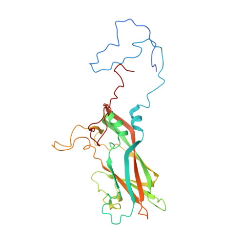
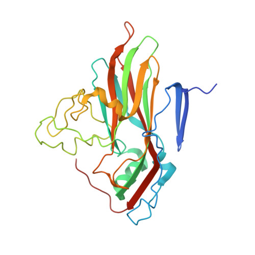
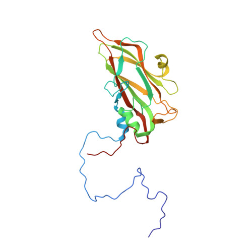
A comparison of the anti-rhinoviral drug binding pocket in HRV14 and HRV1A.

Kim, K.H.Willingmann, P.Gong, Z.X.Kremer, M.J.Chapman, M.S.Minor, I.Oliveira, M.A.Rossmann, M.G.Andries, K.Diana, G.D.Dutko, F.J.McKinlay, M.A.Pevear, D.C.

(1993) J Mol Biol 230: 206-227

  • DOI: https://doi.org/10.1006/jmbi.1993.1137
  • Primary Citation of Related Structures:  
    2HWB, 2HWC, 2HWD, 2HWE, 2HWF

  • PubMed Abstract: 

    The three-dimensional structures of two human rhinovirus serotypes (HRV14 and HRV1A) are compared when complexed with various antiviral agents. Although these agents all bind into the same hydrophobic pocket, the exact viral-drug interactions differ. In the absence of drugs, the pocket is occupied by a fatty acid in HRV1A, but is empty in HRV14 except for two water molecules. The conformation of each drug is dependent upon the shape of the hydrophobic pocket. In HRV14 the major residues determining the shape of the binding site are Y1128, P1174 and M1224, corresponding to I1125, M1169 and I1220 in HRV1A. When there is no cofactor or a drug in the pocket, the entrance to the pocket is open. However, the entrance is closed when the pocket is occupied by a cofactor or a drug. There are relatively small conformational changes when the agents displace the natural cofactor in HRV1A. In contrast, there are much larger conformational changes on binding a drug in HRV14. These differences cause an inhibition of viral attachment in HRV14 but not in HRV1A. Binding of the drugs results in three additional interprotomer hydrogen bonds in HRV14 and one in HRV1A. These hydrogen bonds and a potential loss of flexibility upon efficient packing of the pocket may contribute to the inhibition of uncoating in both serotypes.


  • Organizational Affiliation

    Department of Biological Sciences, Purdue University West Lafayette, IN 47907.


Macromolecules
Find similar proteins by:  (by identity cutoff)  |  3D Structure
Entity ID: 1
MoleculeChains Sequence LengthOrganismDetailsImage
HUMAN RHINOVIRUS 14 COAT PROTEIN (SUBUNIT VP1)A [auth 1]289rhinovirus B14Mutation(s): 0 
UniProt
Find proteins for P03303 (Human rhinovirus 14)
Explore P03303 
Go to UniProtKB:  P03303
Entity Groups  
Sequence Clusters30% Identity50% Identity70% Identity90% Identity95% Identity100% Identity
UniProt GroupP03303
Sequence Annotations
Expand
  • Reference Sequence
Find similar proteins by:  (by identity cutoff)  |  3D Structure
Entity ID: 2
MoleculeChains Sequence LengthOrganismDetailsImage
HUMAN RHINOVIRUS 14 COAT PROTEIN (SUBUNIT VP2)B [auth 2]262rhinovirus B14Mutation(s): 0 
UniProt
Find proteins for P03303 (Human rhinovirus 14)
Explore P03303 
Go to UniProtKB:  P03303
Entity Groups  
Sequence Clusters30% Identity50% Identity70% Identity90% Identity95% Identity100% Identity
UniProt GroupP03303
Sequence Annotations
Expand
  • Reference Sequence
Find similar proteins by:  (by identity cutoff)  |  3D Structure
Entity ID: 3
MoleculeChains Sequence LengthOrganismDetailsImage
HUMAN RHINOVIRUS 14 COAT PROTEIN (SUBUNIT VP3)C [auth 3]236rhinovirus B14Mutation(s): 0 
UniProt
Find proteins for P03303 (Human rhinovirus 14)
Explore P03303 
Go to UniProtKB:  P03303
Entity Groups  
Sequence Clusters30% Identity50% Identity70% Identity90% Identity95% Identity100% Identity
UniProt GroupP03303
Sequence Annotations
Expand
  • Reference Sequence
Find similar proteins by:  (by identity cutoff)  |  3D Structure
Entity ID: 4
MoleculeChains Sequence LengthOrganismDetailsImage
HUMAN RHINOVIRUS 14 COAT PROTEIN (SUBUNIT VP4)D [auth 4]68rhinovirus B14Mutation(s): 0 
UniProt
Find proteins for P03303 (Human rhinovirus 14)
Explore P03303 
Go to UniProtKB:  P03303
Entity Groups  
Sequence Clusters30% Identity50% Identity70% Identity90% Identity95% Identity100% Identity
UniProt GroupP03303
Sequence Annotations
Expand
  • Reference Sequence
Small Molecules
Ligands 1 Unique
IDChains Name / Formula / InChI Key2D Diagram3D Interactions
W54
Query on W54

Download Ideal Coordinates CCD File 
E [auth 1]5-(5-(2,6-DICHLORO-4-(4,5-DIHYDRO-2-OXAZOLY)PHENOXY)PENTYL)-3-METHYL ISOXAZOLE
C18 H20 Cl2 N2 O3
JJDHAOLOHQTGMG-UHFFFAOYSA-N
Experimental Data & Validation

Experimental Data

  • Method: X-RAY DIFFRACTION
  • Resolution: 3.00 Å
  • Space Group: P 21 3
Unit Cell:
Length ( Å )Angle ( ˚ )
a = 445.1α = 90
b = 445.1β = 90
c = 445.1γ = 90

Structure Validation

View Full Validation Report


Entry History 

Deposition Data

Revision History  (Full details and data files)

  • Version 1.0: 1994-11-01
    Type: Initial release
  • Version 1.1: 2008-03-24
    Changes: Version format compliance
  • Version 1.2: 2011-07-13
    Changes: Version format compliance
  • Version 2.0: 2023-01-18
    Type: Remediation
    Changes: Advisory, Atomic model, Data collection, Database references, Derived calculations, Other, Refinement description