4DOE

The liganded structure of Cbescii CelA GH9 module


Experimental Data Snapshot

  • Method: X-RAY DIFFRACTION
  • Resolution: 1.56 Å
  • R-Value Free: 0.175 
  • R-Value Work: 0.148 
  • R-Value Observed: 0.149 

wwPDB Validation   3D Report Full Report


This is version 2.1 of the entry. See complete history


Literature

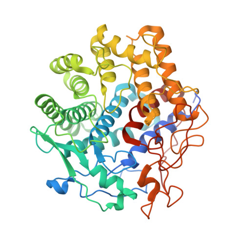
Revealing nature's cellulase diversity: the digestion mechanism of Caldicellulosiruptor bescii CelA.

Brunecky, R.Alahuhta, M.Xu, Q.Donohoe, B.S.Crowley, M.F.Kataeva, I.A.Yang, S.J.Resch, M.G.Adams, M.W.Lunin, V.V.Himmel, M.E.Bomble, Y.J.

(2013) Science 342: 1513-1516

  • DOI: https://doi.org/10.1126/science.1244273
  • Primary Citation of Related Structures:  
    4DOD, 4DOE, 4EL8

  • PubMed Abstract: 

    Most fungi and bacteria degrade plant cell walls by secreting free, complementary enzymes that hydrolyze cellulose; however, some bacteria use large enzymatic assemblies called cellulosomes, which recruit complementary enzymes to protein scaffolds. The thermophilic bacterium Caldicellulosiruptor bescii uses an intermediate strategy, secreting many free cellulases that contain multiple catalytic domains. One of these, CelA, comprises a glycoside hydrolase family 9 and a family 48 catalytic domain, as well as three type III cellulose-binding modules. In the saccharification of a common cellulose standard, Avicel, CelA outperforms mixtures of commercially relevant exo- and endoglucanases. From transmission electron microscopy studies of cellulose after incubation with CelA, we report morphological features that suggest that CelA not only exploits the common surface ablation mechanism driven by general cellulase processivity, but also excavates extensive cavities into the surface of the substrate. These results suggest that nature's repertoire of cellulose digestion paradigms remain only partially discovered and understood.


  • Organizational Affiliation

    Biosciences Center, National Renewable Energy Laboratory, 15013 Denver West Parkway, Golden, CO 80401, USA.


Macromolecules
Find similar proteins by:  (by identity cutoff)  |  3D Structure
Entity ID: 1
MoleculeChains Sequence LengthOrganismDetailsImage
1,4-beta-glucanase475Caldicellulosiruptor besciiMutation(s): 0 
Gene Names: celA
EC: 3.2.1.4
UniProt
Find proteins for P96311 (Caldicellulosiruptor bescii)
Explore P96311 
Go to UniProtKB:  P96311
Entity Groups  
Sequence Clusters30% Identity50% Identity70% Identity90% Identity95% Identity100% Identity
UniProt GroupP96311
Sequence Annotations
Expand
  • Reference Sequence
Oligosaccharides

Help

Entity ID: 2
MoleculeChains Length2D Diagram Glycosylation3D Interactions
beta-D-glucopyranose-(1-4)-alpha-D-glucopyranose
B
2N/A
Glycosylation Resources
GlyTouCan:  G86802XL
GlyCosmos:  G86802XL
Entity ID: 3
MoleculeChains Length2D Diagram Glycosylation3D Interactions
beta-D-glucopyranose-(1-4)-beta-D-glucopyranose-(1-4)-beta-D-glucopyranose
C
3N/A
Glycosylation Resources
GlyTouCan:  G14338VK
GlyCosmos:  G14338VK
GlyGen:  G14338VK
Small Molecules
Ligands 5 Unique
IDChains Name / Formula / InChI Key2D Diagram3D Interactions
SO4
Query on SO4

Download Ideal Coordinates CCD File 
LA [auth A],
MA [auth A],
NA [auth A]
SULFATE ION
O4 S
QAOWNCQODCNURD-UHFFFAOYSA-L
GOL
Query on GOL

Download Ideal Coordinates CCD File 
F [auth A]
G [auth A]
H [auth A]
I [auth A]
J [auth A]
F [auth A],
G [auth A],
H [auth A],
I [auth A],
J [auth A],
K [auth A]
GLYCEROL
C3 H8 O3
PEDCQBHIVMGVHV-UHFFFAOYSA-N
DIO
Query on DIO

Download Ideal Coordinates CCD File 
D [auth A]1,4-DIETHYLENE DIOXIDE
C4 H8 O2
RYHBNJHYFVUHQT-UHFFFAOYSA-N
EDO
Query on EDO

Download Ideal Coordinates CCD File 
AA [auth A]
BA [auth A]
CA [auth A]
DA [auth A]
EA [auth A]
AA [auth A],
BA [auth A],
CA [auth A],
DA [auth A],
EA [auth A],
FA [auth A],
GA [auth A],
HA [auth A],
IA [auth A],
JA [auth A],
KA [auth A],
L [auth A],
M [auth A],
N [auth A],
O [auth A],
P [auth A],
Q [auth A],
R [auth A],
S [auth A],
T [auth A],
U [auth A],
V [auth A],
W [auth A],
X [auth A],
Y [auth A],
Z [auth A]
1,2-ETHANEDIOL
C2 H6 O2
LYCAIKOWRPUZTN-UHFFFAOYSA-N
CA
Query on CA

Download Ideal Coordinates CCD File 
E [auth A]CALCIUM ION
Ca
BHPQYMZQTOCNFJ-UHFFFAOYSA-N
Biologically Interesting Molecules (External Reference) 2 Unique
Experimental Data & Validation

Experimental Data

  • Method: X-RAY DIFFRACTION
  • Resolution: 1.56 Å
  • R-Value Free: 0.175 
  • R-Value Work: 0.148 
  • R-Value Observed: 0.149 
  • Space Group: P 31 2 1
Unit Cell:
Length ( Å )Angle ( ˚ )
a = 147.931α = 90
b = 147.931β = 90
c = 59.098γ = 120
Software Package:
Software NamePurpose
PROTEUM PLUSdata collection
MOLREPphasing
REFMACrefinement
PROTEUM PLUSdata reduction
PROTEUM PLUSdata scaling

Structure Validation

View Full Validation Report


Entry History 

Deposition Data

Revision History  (Full details and data files)

  • Version 1.0: 2013-02-20
    Type: Initial release
  • Version 1.1: 2013-12-18
    Changes: Database references
  • Version 1.2: 2014-01-08
    Changes: Database references
  • Version 2.0: 2020-07-29
    Type: Remediation
    Reason: Carbohydrate remediation
    Changes: Atomic model, Data collection, Database references, Derived calculations, Non-polymer description, Structure summary
  • Version 2.1: 2023-09-13
    Changes: Data collection, Database references, Refinement description, Structure summary