4F8N

X-ray structure of PsaA from Yersinia pestis, in complex with galactose and phosphate choline


Experimental Data Snapshot

  • Method: X-RAY DIFFRACTION
  • Resolution: 2.50 Å
  • R-Value Free: 0.282 
  • R-Value Work: 0.245 
  • R-Value Observed: 0.247 

wwPDB Validation   3D Report Full Report


Ligand Structure Quality Assessment 


This is version 1.5 of the entry. See complete history


Literature

Structural basis for the specific recognition of dual receptors by the homopolymeric pH 6 antigen (Psa) fimbriae of Yersinia pestis.

Bao, R.Nair, M.K.Tang, W.K.Esser, L.Sadhukhan, A.Holland, R.L.Xia, D.Schifferli, D.M.

(2013) Proc Natl Acad Sci U S A 110: 1065-1070

  • DOI: https://doi.org/10.1073/pnas.1212431110
  • Primary Citation of Related Structures:  
    4F8L, 4F8N, 4F8O, 4F8P

  • PubMed Abstract: 

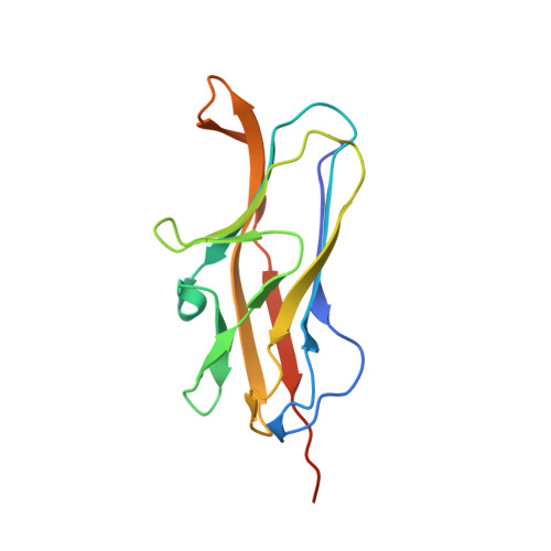
    The pH 6 antigen (Psa) of Yersinia pestis consists of fimbriae that bind to two receptors: β1-linked galactosyl residues in glycosphingolipids and the phosphocholine group in phospholipids. Despite the ubiquitous presence of either moiety on the surface of many mammalian cells, Y. pestis appears to prefer interacting with certain types of human cells, such as macrophages and alveolar epithelial cells of the lung. The molecular mechanism of this apparent selectivity is not clear. Site-directed mutagenesis of the consensus choline-binding motif in the sequence of PsaA, the subunit of the Psa fimbrial homopolymer, identified residues that abolish galactosylceramide binding, phosphatidylcholine binding, or both. The crystal structure of PsaA in complex with both galactose and phosphocholine reveals separate receptor binding sites that share a common structural motif, thus suggesting a potential interaction between the two sites. Mutagenesis of this shared structural motif identified Tyr126, which is part of the choline-binding consensus sequence but is found in direct contact with the galactose in the structure of PsaA, important for both receptor binding. Thus, this structure depicts a fimbrial subunit that forms a polymeric adhesin with a unique arrangement of dual receptor binding sites. These findings move the field forward by providing insights into unique types of multiple receptor-ligand interactions and should steer research into the synthesis of dual receptor inhibitor molecules to slow down the rapid progression of plague.


  • Organizational Affiliation

    Laboratory of Cell Biology, Center for Cancer Research, National Cancer Institute, National Institutes of Health, Bethesda, MD 20892, USA.


Macromolecules
Find similar proteins by:  (by identity cutoff)  |  3D Structure
Entity ID: 1
MoleculeChains Sequence LengthOrganismDetailsImage
pH 6 antigen145Yersinia pestisMutation(s): 0 
Gene Names: psaAy2882YPO1303YP_1289
UniProt
Find proteins for P31522 (Yersinia pestis)
Explore P31522 
Go to UniProtKB:  P31522
Entity Groups  
Sequence Clusters30% Identity50% Identity70% Identity90% Identity95% Identity100% Identity
UniProt GroupP31522
Sequence Annotations
Expand
  • Reference Sequence
Small Molecules
Binding Affinity Annotations 
IDSourceBinding Affinity
GAL Binding MOAD:  4F8N IC50: 4.00e+7 (nM) from 1 assay(s)
Experimental Data & Validation

Experimental Data

  • Method: X-RAY DIFFRACTION
  • Resolution: 2.50 Å
  • R-Value Free: 0.282 
  • R-Value Work: 0.245 
  • R-Value Observed: 0.247 
  • Space Group: P 2 2 21
Unit Cell:
Length ( Å )Angle ( ˚ )
a = 26.154α = 90
b = 54.922β = 90
c = 103.271γ = 90
Software Package:
Software NamePurpose
DENZOdata reduction
SCALEPACKdata scaling
PHASERphasing
REFMACrefinement
PDB_EXTRACTdata extraction
HKL-2000data collection
PHENIXrefinement

Structure Validation

View Full Validation Report


Ligand Structure Quality Assessment 


Entry History 

Deposition Data

Revision History  (Full details and data files)

  • Version 1.0: 2013-05-22
    Type: Initial release
  • Version 1.1: 2013-08-07
    Changes: Database references
  • Version 1.2: 2017-08-16
    Changes: Refinement description, Source and taxonomy
  • Version 1.3: 2017-11-15
    Changes: Refinement description
  • Version 1.4: 2020-07-29
    Type: Remediation
    Reason: Carbohydrate remediation
    Changes: Data collection, Database references, Derived calculations, Structure summary
  • Version 1.5: 2024-02-28
    Changes: Data collection, Database references, Structure summary