5XPS

Crystal Structure of Transketolase in complex with erythrose-4-phosphate from Pichia Stipitis


Experimental Data Snapshot

  • Method: X-RAY DIFFRACTION
  • Resolution: 1.07 Å
  • R-Value Free: 0.132 
  • R-Value Work: 0.119 
  • R-Value Observed: 0.119 

wwPDB Validation   3D Report Full Report


Ligand Structure Quality Assessment 


This is version 1.2 of the entry. See complete history


Literature

Evidence of Diradicals Involved in the Yeast Transketolase Catalyzed Keto-Transferring Reactions.

Hsu, N.S.Wang, Y.L.Lin, K.H.Chang, C.F.Ke, S.C.Lyu, S.Y.Hsu, L.J.Li, Y.S.Chen, S.C.Wang, K.C.Li, T.L.

(2018) Chembiochem 19: 2395-2402

  • DOI: https://doi.org/10.1002/cbic.201800378
  • Primary Citation of Related Structures:  
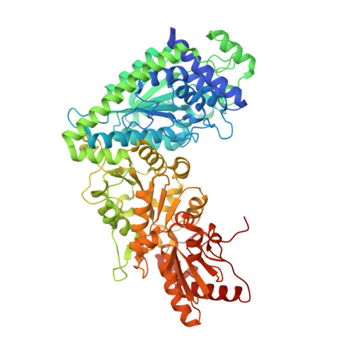
    5XPS, 5XQK, 5XRV, 5XT0, 5XT4, 5XTX, 5XU2, 5XU9

  • PubMed Abstract: 

    Transketolase (TK) catalyzes a reversible transfer of a two-carbon (C 2 ) unit between phosphoketose donors and phosphoaldose acceptors, for which the group-transfer reaction that follows a one- or two-electron mechanism and the force that breaks the C2"-C3" bond of the ketose donors remain unresolved. Herein, we report ultrahigh-resolution crystal structures of a TK (TKps) from Pichia stipitis in previously undiscovered intermediate states and support a diradical mechanism for a reversible group-transfer reaction. In conjunction with MS, NMR spectroscopy, EPR and computational analyses, it is concluded that the enzyme-catalyzed non-Kekulé diradical cofactor brings about the C2"-C3" bond cleavage/formation for the C 2 -unit transfer reaction, for which suppression of activation energy and activation and destabilization of enzymatic intermediates are facilitated.


  • Organizational Affiliation

    Genomics Research Center, Academia Sinica, Taipei, 115, Taiwan.


Macromolecules
Find similar proteins by:  (by identity cutoff)  |  3D Structure
Entity ID: 1
MoleculeChains Sequence LengthOrganismDetailsImage
Transketolase697Scheffersomyces stipitis CBS 6054Mutation(s): 0 
Gene Names: TKTTKT1PICST_67105
EC: 2.2.1.1
UniProt
Find proteins for P34736 (Scheffersomyces stipitis (strain ATCC 58785 / CBS 6054 / NBRC 10063 / NRRL Y-11545))
Explore P34736 
Go to UniProtKB:  P34736
Entity Groups  
Sequence Clusters30% Identity50% Identity70% Identity90% Identity95% Identity100% Identity
UniProt GroupP34736
Sequence Annotations
Expand
  • Reference Sequence
Small Molecules
Ligands 4 Unique
IDChains Name / Formula / InChI Key2D Diagram3D Interactions
TPP
Query on TPP

Download Ideal Coordinates CCD File 
F [auth A]THIAMINE DIPHOSPHATE
C12 H19 N4 O7 P2 S
AYEKOFBPNLCAJY-UHFFFAOYSA-O
E4P
Query on E4P

Download Ideal Coordinates CCD File 
G [auth A]ERYTHOSE-4-PHOSPHATE
C4 H9 O7 P
NGHMDNPXVRFFGS-IUYQGCFVSA-N
PEG
Query on PEG

Download Ideal Coordinates CCD File 
B [auth A],
C [auth A],
D [auth A],
E [auth A]
DI(HYDROXYETHYL)ETHER
C4 H10 O3
MTHSVFCYNBDYFN-UHFFFAOYSA-N
CA
Query on CA

Download Ideal Coordinates CCD File 
H [auth A]CALCIUM ION
Ca
BHPQYMZQTOCNFJ-UHFFFAOYSA-N
Experimental Data & Validation

Experimental Data

  • Method: X-RAY DIFFRACTION
  • Resolution: 1.07 Å
  • R-Value Free: 0.132 
  • R-Value Work: 0.119 
  • R-Value Observed: 0.119 
  • Space Group: C 2 2 21
Unit Cell:
Length ( Å )Angle ( ˚ )
a = 101.149α = 90
b = 185.246β = 90
c = 98.71γ = 90
Software Package:
Software NamePurpose
REFMACrefinement
HKL-2000data scaling

Structure Validation

View Full Validation Report


Ligand Structure Quality Assessment 


Entry History 

Deposition Data

Revision History  (Full details and data files)

  • Version 1.0: 2018-06-06
    Type: Initial release
  • Version 1.1: 2018-12-19
    Changes: Data collection, Database references
  • Version 1.2: 2023-11-22
    Changes: Data collection, Database references, Refinement description