5YCL

Crystal structure of HigBA complex from Shigella flexneri


Experimental Data Snapshot

  • Method: X-RAY DIFFRACTION
  • Resolution: 3.10 Å
  • R-Value Free: 0.303 
  • R-Value Work: 0.268 
  • R-Value Observed: 0.272 

wwPDB Validation   3D Report Full Report


This is version 1.1 of the entry. See complete history


Literature

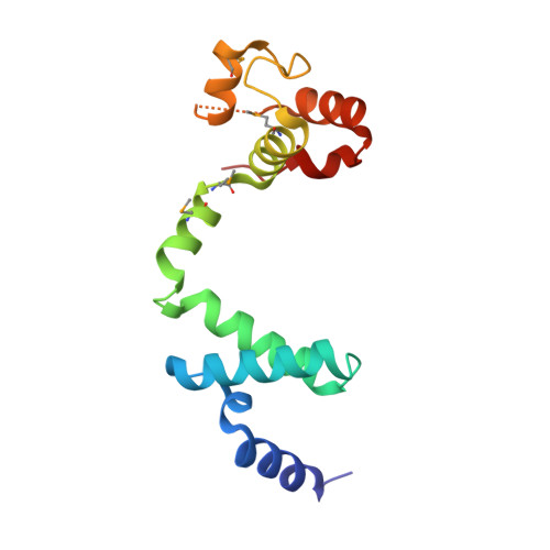
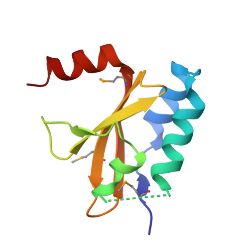
Structural changes of antitoxin HigA from Shigella flexneri by binding of its cognate toxin HigB.

Yoon, W.S.Seok, S.H.Won, H.S.Cho, T.Lee, S.J.Seo, M.D.

(2019) Int J Biol Macromol 130: 99-108

  • DOI: https://doi.org/10.1016/j.ijbiomac.2019.02.111
  • Primary Citation of Related Structures:  
    5YCL, 6IRP

  • PubMed Abstract: 

    In toxin-antitoxin systems, many antitoxin proteins that neutralize their cognate toxin proteins also bind to DNA to repress transcription, and the DNA-binding affinity of the antitoxin is affected by its toxin. We solved crystal structures of the antitoxin HigA (apo- Sf HigA) and its complex with the toxin HigB ( Sf HigBA) from Shigella flexneri. The apo- Sf HigA shows a distinctive V-shaped homodimeric conformation with sequestered N-domains having a novel fold. Sf HigBA appears as a heterotetramer formed by N-terminal dimerization of Sf HigB-bound Sf HigA molecules. The conformational change in Sf HigA upon Sf HigB binding is mediated by rigid-body movements of its C-domains, which accompanied an overall conformational change from wide V-shaped to narrow V-shaped dimer. Consequently, the two putative DNA-binding helices (α7 in each subunit) are repositioned to a conformation more compatible with canonical homodimeric DNA-binding proteins containing HTH motifs. Collectively, this study demonstrates a conformational change in an antitoxin protein, which occurs upon toxin binding and is responsible for regulating antitoxin DNA binding.


  • Organizational Affiliation

    Department of Molecular Science and Technology, Ajou University, Suwon, Gyeonggi 16499, Republic of Korea; College of Pharmacy and Research Institute of Pharmaceutical Science and Technology (RIPST), Ajou University, Suwon, Gyeonggi 16499, Republic of Korea.


Macromolecules
Find similar proteins by:  (by identity cutoff)  |  3D Structure
Entity ID: 1
MoleculeChains Sequence LengthOrganismDetailsImage
Antitoxin HigA
A, C
138Shigella flexneriMutation(s): 0 
Gene Names: higASF3122S3329
UniProt
Find proteins for P67703 (Shigella flexneri)
Explore P67703 
Go to UniProtKB:  P67703
Entity Groups  
Sequence Clusters30% Identity50% Identity70% Identity90% Identity95% Identity100% Identity
UniProt GroupP67703
Sequence Annotations
Expand
  • Reference Sequence
Find similar proteins by:  (by identity cutoff)  |  3D Structure
Entity ID: 2
MoleculeChains Sequence LengthOrganismDetailsImage
mRNA interferase HigB
B, D
101Shigella flexneriMutation(s): 0 
Gene Names: higBSF3123S3330
EC: 3.1
UniProt
Find proteins for P64580 (Shigella flexneri)
Explore P64580 
Go to UniProtKB:  P64580
Entity Groups  
Sequence Clusters30% Identity50% Identity70% Identity90% Identity95% Identity100% Identity
UniProt GroupP64580
Sequence Annotations
Expand
  • Reference Sequence
Small Molecules
Modified Residues  1 Unique
IDChains TypeFormula2D DiagramParent
MSE
Query on MSE
A, C
L-PEPTIDE LINKINGC5 H11 N O2 SeMET
Experimental Data & Validation

Experimental Data

  • Method: X-RAY DIFFRACTION
  • Resolution: 3.10 Å
  • R-Value Free: 0.303 
  • R-Value Work: 0.268 
  • R-Value Observed: 0.272 
  • Space Group: P 31 2 1
Unit Cell:
Length ( Å )Angle ( ˚ )
a = 78.209α = 90
b = 78.209β = 90
c = 183.824γ = 120
Software Package:
Software NamePurpose
PHENIXrefinement
PHENIXphasing
PHENIXdata processing
HKL-2000data scaling

Structure Validation

View Full Validation Report


Entry History 

Deposition Data

Revision History  (Full details and data files)

  • Version 1.0: 2018-09-19
    Type: Initial release
  • Version 1.1: 2019-09-04
    Changes: Data collection, Database references