6CMK

Crystal structure of Citrobacter koseri AztD


Experimental Data Snapshot

  • Method: X-RAY DIFFRACTION
  • Resolution: 1.73 Å
  • R-Value Free: 0.188 
  • R-Value Work: 0.153 
  • R-Value Observed: 0.154 

wwPDB Validation   3D Report Full Report


This is version 1.2 of the entry. See complete history


Literature

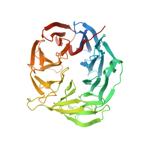
Crystal structures of AztD provide mechanistic insights into direct zinc transfer between proteins.

Neupane, D.P.Fullam, S.H.Chacon, K.N.Yukl, E.T.

(2019) Commun Biol 2: 308-308

  • DOI: https://doi.org/10.1038/s42003-019-0542-z
  • Primary Citation of Related Structures:  
    6CK1, 6CMK, 6N01

  • PubMed Abstract: 

    Zinc acquisition from limited environments is critical for bacterial survival and pathogenesis. AztD has been identified as a periplasmic or cell surface zinc-binding protein in numerous bacterial species. In Paracoccus denitrificans , AztD can transfer zinc directly to AztC, the solute binding protein for a zinc-specific ATP-binding cassette transporter system, suggesting a role in zinc acquisition and homeostasis. Here, we present the first cry stal structures of AztD from P. denitrificans and tbe human pathogen Citrobacter koseri , revealing a beta-propeller fold and two high-affinity zinc-binding sites that are highly conserved among AztD homologs. These structures combined with transfer assays using WT and mutant proteins provide rare insight into the mechanism of direct zinc transfer from one protein to another. Given the importance of zinc import to bacterial pathogenesis, these insights may prove valuable to the development of zinc transfer inhibitors as antibiotics.


  • Organizational Affiliation

    1Department of Chemistry and Biochemistry, New Mexico State University, Las Cruces, NM 88003 USA.


Macromolecules
Find similar proteins by:  (by identity cutoff)  |  3D Structure
Entity ID: 1
MoleculeChains Sequence LengthOrganismDetailsImage
AztD proteinA [auth B],
B [auth A]
421Citrobacter koseri ATCC BAA-895Mutation(s): 0 
Gene Names: CKO_00948
UniProt
Find proteins for A8AF35 (Citrobacter koseri (strain ATCC BAA-895 / CDC 4225-83 / SGSC4696))
Explore A8AF35 
Go to UniProtKB:  A8AF35
Entity Groups  
Sequence Clusters30% Identity50% Identity70% Identity90% Identity95% Identity100% Identity
UniProt GroupA8AF35
Sequence Annotations
Expand
  • Reference Sequence
Small Molecules
Ligands 5 Unique
IDChains Name / Formula / InChI Key2D Diagram3D Interactions
MES
Query on MES

Download Ideal Coordinates CCD File 
C [auth B],
H [auth A]
2-(N-MORPHOLINO)-ETHANESULFONIC ACID
C6 H13 N O4 S
SXGZJKUKBWWHRA-UHFFFAOYSA-N
PGE
Query on PGE

Download Ideal Coordinates CCD File 
M [auth A]TRIETHYLENE GLYCOL
C6 H14 O4
ZIBGPFATKBEMQZ-UHFFFAOYSA-N
PEG
Query on PEG

Download Ideal Coordinates CCD File 
N [auth A]DI(HYDROXYETHYL)ETHER
C4 H10 O3
MTHSVFCYNBDYFN-UHFFFAOYSA-N
ZN
Query on ZN

Download Ideal Coordinates CCD File 
D [auth B],
E [auth B],
I [auth A],
J [auth A]
ZINC ION
Zn
PTFCDOFLOPIGGS-UHFFFAOYSA-N
EDO
Query on EDO

Download Ideal Coordinates CCD File 
F [auth B],
G [auth B],
K [auth A],
L [auth A]
1,2-ETHANEDIOL
C2 H6 O2
LYCAIKOWRPUZTN-UHFFFAOYSA-N
Experimental Data & Validation

Experimental Data

  • Method: X-RAY DIFFRACTION
  • Resolution: 1.73 Å
  • R-Value Free: 0.188 
  • R-Value Work: 0.153 
  • R-Value Observed: 0.154 
  • Space Group: P 1 21 1
Unit Cell:
Length ( Å )Angle ( ˚ )
a = 54.23α = 90
b = 128.79β = 105.2
c = 56.85γ = 90
Software Package:
Software NamePurpose
PHENIXrefinement
MOSFLMdata reduction
Aimlessdata scaling
PHENIXphasing

Structure Validation

View Full Validation Report


Entry History & Funding Information

Deposition Data

  • Released Date: 2019-03-13 
  • Deposition Author(s): Yukl, E.T.

Funding OrganizationLocationGrant Number
National Institutes of Health/National Institute of General Medical Sciences (NIH/NIGMS)United StatesSC2GM111170

Revision History  (Full details and data files)

  • Version 1.0: 2019-03-13
    Type: Initial release
  • Version 1.1: 2019-09-25
    Changes: Data collection, Database references
  • Version 1.2: 2020-01-01
    Changes: Author supporting evidence