6HHZ

Klebsiella pneumoniae Seryl-tRNA Synthetase in Complex with 5'-O-(N-(L-seryl)-sulfamoyl)cytidine


Experimental Data Snapshot

  • Method: X-RAY DIFFRACTION
  • Resolution: 2.15 Å
  • R-Value Free: 0.226 
  • R-Value Work: 0.200 
  • R-Value Observed: 0.201 

wwPDB Validation   3D Report Full Report


Ligand Structure Quality Assessment 


This is version 1.4 of the entry. See complete history


Literature

Structural Insights into the Binding of Natural Pyrimidine-Based Inhibitors of Class II Aminoacyl-tRNA Synthetases.

Pang, L.Nautiyal, M.De Graef, S.Gadakh, B.Zorzini, V.Economou, A.Strelkov, S.V.Van Aerschot, A.Weeks, S.D.

(2020) ACS Chem Biol 15: 407-415

  • DOI: https://doi.org/10.1021/acschembio.9b00887
  • Primary Citation of Related Structures:  
    6HDZ, 6HE1, 6HE3, 6HHV, 6HHW, 6HHX, 6HHY, 6HHZ, 6HI0, 6RLT, 6RLU, 6RLV, 6S30, 6SJC

  • PubMed Abstract: 

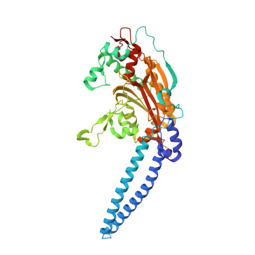
    The pyrimidine-containing Trojan horse antibiotics albomycin and a recently discovered cytidine-containing microcin C analog target the class II seryl- and aspartyl-tRNA synthetases (serRS and aspRS), respectively. The active components of these compounds are competitive inhibitors that mimic the aminoacyl-adenylate intermediate. How they effectively substitute for the interactions mediated by the canonical purine group is unknown. Employing nonhydrolyzable aminoacyl-sulfamoyl nucleosides substituting the base with cytosine, uracil, and N3-methyluracil the structure-activity relationship of the natural compounds was evaluated. In vitro using E. coli serRS and aspRS, the best compounds demonstrated IC 50 values in the low nanomolar range, with a clear preference for cytosine or N3-methyluracil over uracil. X-ray crystallographic structures of K. pneumoniae serRS and T. thermophilus aspRS in complex with the compounds showed the contribution of structured waters and residues in the conserved motif-2 loop in defining base preference. Utilizing the N3-methyluracil bound serRS structure, MD simulations of the fully modified albomycin base were performed to identify the interacting network that drives stable association. This analysis pointed to key interactions with a methionine in the motif-2 loop. Interestingly, this residue is mutated to a glycine in a second serRS (serRS2) found in albomycin-producing actinobacteria possessing self-immunity to this antibiotic. A comparative study demonstrated that serRS2 is poorly inhibited by the pyrimidine-containing intermediate analogs, and an equivalent mutation in E. coli serRS significantly decreased the affinity of the cytosine congener. These findings highlight the crucial role of dynamics and solvation of the motif-2 loop in modulating the binding of the natural antibiotics.


  • Organizational Affiliation

    Biocrystallography, Department of Pharmaceutical and Pharmacological Sciences , KU Leuven , Herestraat 49 Box 822 , B-3000 Leuven , Belgium.


Macromolecules
Find similar proteins by:  (by identity cutoff)  |  3D Structure
Entity ID: 1
MoleculeChains Sequence LengthOrganismDetailsImage
Serine--tRNA ligase430Klebsiella pneumoniaeMutation(s): 0 
Gene Names: serSKPN78578_09000KPN_00925
EC: 6.1.1.11
UniProt
Find proteins for A6T6Z0 (Klebsiella pneumoniae subsp. pneumoniae (strain ATCC 700721 / MGH 78578))
Explore A6T6Z0 
Go to UniProtKB:  A6T6Z0
Entity Groups  
Sequence Clusters30% Identity50% Identity70% Identity90% Identity95% Identity100% Identity
UniProt GroupA6T6Z0
Sequence Annotations
Expand
  • Reference Sequence
Small Molecules
Ligands 5 Unique
IDChains Name / Formula / InChI Key2D Diagram3D Interactions
FZQ (Subject of Investigation/LOI)
Query on FZQ

Download Ideal Coordinates CCD File 
E [auth A]5'-O-(N-(L-seryl)-sulfamoyl)cytidine
C12 H19 N5 O9 S
KLHALWQAANSHJB-DANLAGSESA-N
1PE
Query on 1PE

Download Ideal Coordinates CCD File 
J [auth A]PENTAETHYLENE GLYCOL
C10 H22 O6
JLFNLZLINWHATN-UHFFFAOYSA-N
EDO
Query on EDO

Download Ideal Coordinates CCD File 
B [auth A],
C [auth A],
D [auth A]
1,2-ETHANEDIOL
C2 H6 O2
LYCAIKOWRPUZTN-UHFFFAOYSA-N
CA
Query on CA

Download Ideal Coordinates CCD File 
F [auth A],
G [auth A],
H [auth A]
CALCIUM ION
Ca
BHPQYMZQTOCNFJ-UHFFFAOYSA-N
CL
Query on CL

Download Ideal Coordinates CCD File 
I [auth A]CHLORIDE ION
Cl
VEXZGXHMUGYJMC-UHFFFAOYSA-M
Experimental Data & Validation

Experimental Data

  • Method: X-RAY DIFFRACTION
  • Resolution: 2.15 Å
  • R-Value Free: 0.226 
  • R-Value Work: 0.200 
  • R-Value Observed: 0.201 
  • Space Group: P 43 21 2
Unit Cell:
Length ( Å )Angle ( ˚ )
a = 83.351α = 90
b = 83.351β = 90
c = 229.329γ = 90
Software Package:
Software NamePurpose
XDSdata reduction
Aimlessdata scaling
BUSTERrefinement
PDB_EXTRACTdata extraction
PHASERphasing

Structure Validation

View Full Validation Report


Ligand Structure Quality Assessment 


Entry History & Funding Information

Deposition Data


Funding OrganizationLocationGrant Number
Research Foundation - FlandersBelgiumG077814N
Research Foundation - FlandersBelgiumG0A4616N
Research Foundation - FlandersBelgium1S53516N

Revision History  (Full details and data files)

  • Version 1.0: 2019-12-18
    Type: Initial release
  • Version 1.1: 2020-01-08
    Changes: Database references
  • Version 1.2: 2020-01-15
    Changes: Database references
  • Version 1.3: 2020-03-04
    Changes: Database references
  • Version 1.4: 2024-01-17
    Changes: Data collection, Database references, Derived calculations, Refinement description