6IZI

Crystal structure of E. coli peptide deformylase and methionine aminopeptidase fitted into the cryo-EM density map of the complex


Experimental Data Snapshot

  • Method: ELECTRON MICROSCOPY
  • Resolution: 11.8 Å
  • Aggregation State: PARTICLE 
  • Reconstruction Method: SINGLE PARTICLE 

wwPDB Validation   3D Report Full Report


This is version 1.1 of the entry. See complete history


Literature

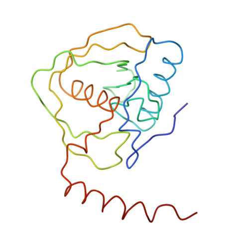
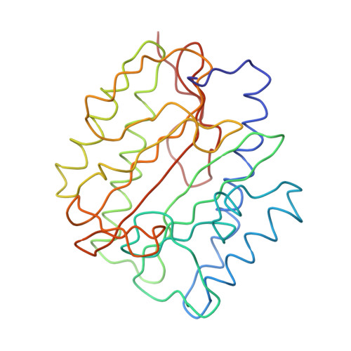
Cryo-EM Structures Reveal Relocalization of MetAP in the Presence of Other Protein Biogenesis Factors at the Ribosomal Tunnel Exit.

Bhakta, S.Akbar, S.Sengupta, J.

(2019) J Mol Biol 431: 1426-1439

  • DOI: https://doi.org/10.1016/j.jmb.2019.02.002
  • Primary Citation of Related Structures:  
    6IY7, 6IZ7, 6IZI, 6J0A, 6J45

  • PubMed Abstract: 

    During protein biosynthesis in bacteria, one of the earliest events that a nascent polypeptide chain goes through is the co-translational enzymatic processing. The event includes two enzymatic pathways: deformylation of the N-terminal methionine by the enzyme peptide deformylase (PDF), followed by methionine excision catalyzed by methionine aminopeptidase (MetAP). During the enzymatic processing, the emerging nascent protein likely remains shielded by the ribosome-associated chaperone trigger factor. The ribosome tunnel exit serves as a stage for recruiting proteins involved in maturation processes of the nascent chain. Co-translational processing of nascent chains is a critical step for subsequent folding and functioning of mature proteins. Here, we present cryo-electron microscopy structures of Escherichia coli (E. coli) ribosome in complex with the nascent chain processing proteins. The structures reveal overlapping binding sites for PDF and MetAP when they bind individually at the tunnel exit site, where L22-L32 protein region provides primary anchoring sites for both proteins. In the absence of PDF, trigger factor can access ribosomal tunnel exit when MetAP occupies its primary binding site. Interestingly, however, in the presence of PDF, when MetAP's primary binding site is already engaged, MetAP has a remarkable ability to occupy an alternative binding site adjacent to PDF. Our study, thus, discloses an unexpected mechanism that MetAP adopts for context-specific ribosome association.


  • Organizational Affiliation

    Structural Biology & Bio-Informatics Division, CSIR-Indian Institute of Chemical Biology, 4, Raja S.C. Mullick Road, Kolkata 700 032, India.


Macromolecules
Find similar proteins by:  (by identity cutoff)  |  3D Structure
Entity ID: 1
MoleculeChains Sequence LengthOrganismDetailsImage
Peptide deformylaseA [auth P]169Escherichia coli K-12Mutation(s): 0 
Gene Names: deffmsb3287JW3248
EC: 3.5.1.88
UniProt
Find proteins for P0A6K3 (Escherichia coli (strain K12))
Explore P0A6K3 
Go to UniProtKB:  P0A6K3
Entity Groups  
Sequence Clusters30% Identity50% Identity70% Identity90% Identity95% Identity100% Identity
UniProt GroupP0A6K3
Sequence Annotations
Expand
  • Reference Sequence
Find similar proteins by:  (by identity cutoff)  |  3D Structure
Entity ID: 2
MoleculeChains Sequence LengthOrganismDetailsImage
Methionine aminopeptidaseB [auth Q]264Escherichia coli K-12Mutation(s): 1 
Gene Names: mapb0168JW0163
EC: 3.4.11.18
UniProt
Find proteins for P0AE18 (Escherichia coli (strain K12))
Explore P0AE18 
Go to UniProtKB:  P0AE18
Entity Groups  
Sequence Clusters30% Identity50% Identity70% Identity90% Identity95% Identity100% Identity
UniProt GroupP0AE18
Sequence Annotations
Expand
  • Reference Sequence
Experimental Data & Validation

Experimental Data

  • Method: ELECTRON MICROSCOPY
  • Resolution: 11.8 Å
  • Aggregation State: PARTICLE 
  • Reconstruction Method: SINGLE PARTICLE 
EM Software:
TaskSoftware PackageVersion
RECONSTRUCTIONSPIDER

Structure Validation

View Full Validation Report


Entry History & Funding Information

Deposition Data


Funding OrganizationLocationGrant Number
Council of Scientific & Industrial ResearchIndia--
Department of Science & Technology (India)IndiaSB/SO/BB-0025/2014

Revision History  (Full details and data files)

  • Version 1.0: 2019-04-17
    Type: Initial release
  • Version 1.1: 2024-03-27
    Changes: Data collection, Database references, Refinement description