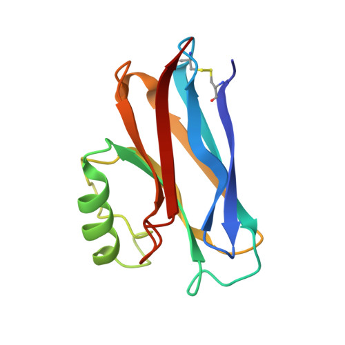
Structure of apo-azurin from Alcaligenes denitrificans at 1.8 A resolution.
Shepard, W.E., Kingston, R.L., Anderson, B.F., Baker, E.N.(1993) Acta Crystallogr D Biol Crystallogr 49: 331-343
- PubMed: 15299522
- DOI: https://doi.org/10.1107/S0907444992013544
- Primary Citation of Related Structures:
1AIZ, 1AZB, 1AZC - PubMed Abstract:
The structure of apo-azurin from Alcaligenes denitrificans has been determined at high resolution by X-ray crystallography. Two separate structure analyses have been carried out, (i) on crystals obtained from solutions of apo-azurin and (ii) on crystals obtained by removal of copper from previously formed crystals of holo-azurin. Data to 1.8 A resolution were collected from the apo-azurin crystals, by Weissenberg photography (with image plates) using synchrotron radiation and by diffractometry, and the structure was refined by restrained least-squares methods to a final R value of 0.160 for all data in the range 10.0-1.8 A. The final model of 1954 protein atoms, 246 water molecules (66 half-weighted), four SO(4)(2-) ions, and two low-occupancy (0.13 and 0.15) Cu atoms has r.m.s. deviations of 0.012, 0.045 and 0.013 A from standard bond lengths, angle distances and planar groups. For copper-removed azurin, data to 2.2 A were collected by diffractometry and the structure refined by restrained least squares to a final R value of 0.158 for all data in the range 10.0-2.2 A. The final model of 1954 protein atoms, 264 water molecules, two SO(4)(2-) ions, two low occupancy (0.18 and 0.22) metal atoms and one unidentified atom (modelled as S) has r.m.s. deviations of 0.013, 0.047 and 0.012 A from standard bond lengths, angle distances and planar groups. The two structures are essentially identical to each other and show no significant differences from the oxidized and reduced holo-azurin structures. The ligand side chains move slightly closer together following the removal of copper, with the radius of the cavity between the three strongly binding ligands, His 46, His 117 and Cys 112, shrinking from 1.31 A in reduced azurin to 1.24 A in oxidized azurin and 1.16 A in apo-azurin. There is a suggestion of increased flexibility in one of the copper-binding loops but the structure supports the view that the copper site found in holo-azurin is a stable structure, defined by the constraints of the polypeptide structure even in the absence of a bound metal ion.
Organizational Affiliation:
Department of Chemistry and Biochemistry, Massey University, Palmerston North, New Zealand.