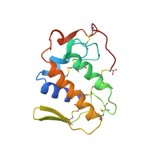
Structure of a calcium-independent phospholipase-like myotoxic protein from Bothrops asper venom.
Arni, R.K., Ward, R.J., Gutierrez, J.M., Tulinsky, A.(1995) Acta Crystallogr D Biol Crystallogr 51: 311-317
- PubMed: 15299297
- DOI: https://doi.org/10.1107/S0907444994011455
- Primary Citation of Related Structures:
1CLP - PubMed Abstract:
Myotoxin II, a myotoxic calcium-independent phospholipase-like protein isolated from the venom of Bothrops asper, possesses no detectable phospholipase activity. The crystal structure has been determined and refined at 2.8 A to an R-factor of 16.5% (F > 3sigma) with excellent stereochemistry. Amino-acid differences between catalytically active phospholipases and myotoxin II in the Ca(2+)-binding region, specifically the substitutions Tyr28-->Asn, Gly32-->Leu and Asp49-->Lys, result in an altered local conformation. The key difference is that the epsilon-amino group of Lys49 fills the site normally occupied by the calcium ion in catalytically active phospholipases. In contrast to the homologous monomeric Lys49 variant from Agkistrodon piscivorus piscivorus, myotoxin II is present as a dimer both in solution and in the crystalline state. The two molecules in the asymmetric unit are related by a nearly perfect twofold axis, yet the dimer is radically different from the dimer formed by the phospholipase from Crotalus atrox. Whereas in C. atrox the dimer interface occludes the active sites, in myotoxin II they are exposed to solvent.
Organizational Affiliation:
Department of Physics, UNEXSP-IBILCE, São José do Rio Preto-SP, Brazil.