1GMK

GRAMICIDIN/KSCN COMPLEX


Experimental Data Snapshot

  • Method: X-RAY DIFFRACTION
  • Resolution: 2.50 Å
  • R-Value Work: 0.192 
  • R-Value Observed: 0.192 

wwPDB Validation   3D Report Full Report


This is version 2.1 of the entry. See complete history


Literature

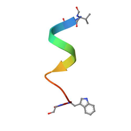
Crystal Structure of the Gramicidin/Potassium Thiocyanate Complex.

Doyle, D.A.Wallace, B.A.

(1997) J Mol Biol 266: 963

  • DOI: https://doi.org/10.1006/jmbi.1996.0837

  • PubMed Abstract: 

    The hydrophobic channel-forming polypeptide gramicidin adopts a left-handed antiparallel double helix conformation with 6.4 residues per turn when in complex with monovalent cation salts in a methanol environment. The crystal structure of the gramicidin/potassium thiocyanate complex (a = 32.06 A, b = 51.80 A, and c = 31.04 A; space group P2(1)2(1)2(1)) has been solved to 2.5 A with an R-factor of 0.193. In the structure, binding sites for the cations are formed by the polypeptide backbone carbonyl groups tilting away from the helix axis toward the ions located in the central lumen. The polypeptide backbone conformations and the side-chain orientations in this potassium complex are significantly different from those in the previously solved gramicidin/caesium chloride crystal complex, due to the requirements for interactions with the smaller sized potassium cation. The locations and numbers of potassium binding sites also differ considerably from the locations and numbers of caesium binding sites in the other structure. Combining information from all the cation binding sites in the two gramicidin/ion complexes produces different views of the three-dimensional structures of a cation as it is transported along a transmembrane pore, and provides an experimental structural basis for modeling the dynamics of peptide-ion binding and ion transport.


  • Organizational Affiliation

    Department of Crystallography, Birkbeck College, University of London, UK.


Macromolecules

Find similar proteins by:  Sequence   |   3D Structure  

Entity ID: 1
MoleculeChains Sequence LengthOrganismDetailsImage
GRAMICIDIN A
A, B, C, D
16Brevibacillus brevisMutation(s): 0 
Entity Groups  
Sequence Clusters30% Identity50% Identity70% Identity90% Identity95% Identity100% Identity
Sequence Annotations
Expand
  • Reference Sequence
Small Molecules
Ligands 3 Unique
IDChains Name / Formula / InChI Key2D Diagram3D Interactions
SCN
Query on SCN

Download Ideal Coordinates CCD File 
F [auth A],
G [auth A],
O [auth B],
V [auth C]
THIOCYANATE ION
C N S
ZMZDMBWJUHKJPS-UHFFFAOYSA-M
K
Query on K

Download Ideal Coordinates CCD File 
CA [auth D],
E [auth A],
N [auth B],
U [auth C]
POTASSIUM ION
K
NPYPAHLBTDXSSS-UHFFFAOYSA-N
MOH
Query on MOH

Download Ideal Coordinates CCD File 
AA [auth C]
BA [auth C]
DA [auth D]
EA [auth D]
FA [auth D]
AA [auth C],
BA [auth C],
DA [auth D],
EA [auth D],
FA [auth D],
GA [auth D],
H [auth A],
HA [auth D],
I [auth A],
IA [auth D],
J [auth A],
JA [auth D],
K [auth A],
KA [auth D],
L [auth A],
M [auth A],
P [auth B],
Q [auth B],
R [auth B],
S [auth B],
T [auth B],
W [auth C],
X [auth C],
Y [auth C],
Z [auth C]
METHANOL
C H4 O
OKKJLVBELUTLKV-UHFFFAOYSA-N
Modified Residues  1 Unique
IDChains TypeFormula2D DiagramParent
FVA
Query on FVA
A, B, C, D
L-PEPTIDE LINKINGC6 H11 N O3VAL
Biologically Interesting Molecules (External Reference) 1 Unique
Experimental Data & Validation

Experimental Data

  • Method: X-RAY DIFFRACTION
  • Resolution: 2.50 Å
  • R-Value Work: 0.192 
  • R-Value Observed: 0.192 
  • Space Group: P 21 21 21
Unit Cell:
Length ( Å )Angle ( ˚ )
a = 32.06α = 90
b = 51.8β = 90
c = 31.04γ = 90
Software Package:
Software NamePurpose
X-PLORmodel building
X-PLORrefinement
CADRALdata reduction
SHELX76data scaling
X-PLORphasing

Structure Validation

View Full Validation Report


Entry History 

Deposition Data

Revision History  (Full details and data files)

  • Version 1.0: 1998-07-01
    Type: Initial release
  • Version 1.1: 2011-06-14
    Changes: Version format compliance
  • Version 1.2: 2011-07-13
    Changes: Version format compliance
  • Version 1.3: 2011-07-27
    Changes: Atomic model, Database references, Derived calculations, Non-polymer description, Structure summary
  • Version 1.4: 2012-12-12
    Changes: Other
  • Version 1.5: 2018-04-18
    Changes: Data collection, Other
  • Version 2.0: 2023-11-15
    Changes: Atomic model, Data collection, Database references, Derived calculations
  • Version 2.1: 2024-04-03
    Changes: Refinement description