Mutagenesis of the histidine ligand in human lactoferrin: iron binding properties and crystal structure of the histidine-253-->methionine mutant.
Nicholson, H., Anderson, B.F., Bland, T., Shewry, S.C., Tweedie, J.W., Baker, E.N.(1997) Biochemistry 36: 341-346
- PubMed: 9003186
- DOI: https://doi.org/10.1021/bi961908y
- Primary Citation of Related Structures:
1HSE - PubMed Abstract:
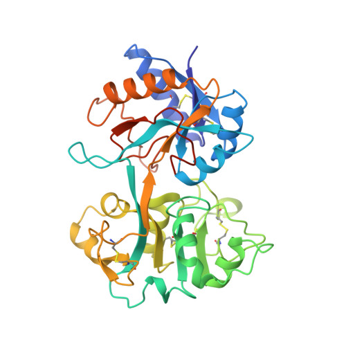
The contribution of the conserved His ligand to iron binding in transferrins has been addressed by site-directed mutagenesis and X-ray crystallographic analysis. His 253 in the N-terminal half-molecule of human lactoferrin, LfN (residues 1-333), has been changed to Gly, Ala, Pro, Thr, Leu, Phe, Met, Tyr, Glu, Gln, and Cys by oligonucleotide-directed mutagenesis. The proteins have been expressed in baby hamster kidney cells, at high levels, and purified. The results show that the His ligand is essential for the stability of the iron binding site. All of the substitutions destabilized iron binding irrespective of whether the replacements were potential iron ligands or not. Iron was lost below pH approximately 6 for the Cys, Glu, and Tyr mutants and below pH 7 or higher for the others, compared with pH 5.0 for LfN. The destabilization is attributed to both steric and electronic effects. The importance of electronic effects has been shown by the crystal structure of the H253M mutant, which has been determined at an effective resolution of 2.5 A and refined to a final R factor of 0.173. The iron atom is changed from six-coordinate to five-coordinate; the Met 253 side chain is not bound to iron even though there appears to be no steric barrier. This is attributed to the poorer affinity of the thioether ligand for Fe(III) compared with imidazole nitrogen. The decreased stability of the iron binding is attributed solely to the loss of the His ligand as the protein conformation and interdomain interactions are unchanged.
Organizational Affiliation:
Department of Biochemistry, Massey University, Palmerston North, New Zealand.