1IZ2

Interactions causing the kinetic trap in serpin protein folding


Experimental Data Snapshot

  • Method: X-RAY DIFFRACTION
  • Resolution: 2.20 Å
  • R-Value Free: 0.278 
  • R-Value Work: 0.211 
  • R-Value Observed: 0.221 

wwPDB Validation   3D Report Full Report


This is version 2.2 of the entry. See complete history


Literature

Interactions causing the kinetic trap in serpin protein folding

Im, H.Woo, M.-S.Hwang, K.Y.Yu, M.-H.

(2002) J Biol Chem 277: 46347-46354

  • DOI: https://doi.org/10.1074/jbc.M207682200
  • Primary Citation of Related Structures:  
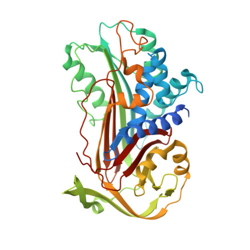
    1IZ2

  • PubMed Abstract: 

    Conformational transition is fundamental to the mechanism of functional regulation in proteins, and serpins (serine protease inhibitors) can provide insight into this process. Serpins are metastable in their native forms, and they ordinarily undergo conformational transition to a stable state only when they form a tight complex with target proteases. The metastable native form is thus considered to be a kinetically trapped folding intermediate. We sought to understand the nature of the serpin kinetic trap as a step toward discovering how conformational transition is regulated. We found that mutations of the B/C beta-barrel of native alpha(1)-antitrypsin, a prototypical serpin, allowed conversion of the molecule into a more stable state. A 2.2 A resolution crystal structure of the stable form (PDB code, ) showed that the reactive site loop is inserted into an A beta-sheet, as in the latent plasminogen activator inhibitor-1. Mutational analyses suggest strongly that interactions not found in the final stable form cause the kinetic trap in serpin protein folding.


  • Organizational Affiliation

    National Creative Research Initiatives, Protein Strain Research Center, Korea Institute of Science and Technology, 39-1 Hawolgok-dong, Sungbuk-gu, Seoul 136-791, Korea.


Macromolecules
Find similar proteins by:  (by identity cutoff)  |  3D Structure
Entity ID: 1
MoleculeChains Sequence LengthOrganismDetailsImage
alpha1-antitrypsin394Homo sapiensMutation(s): 10 
UniProt & NIH Common Fund Data Resources
Find proteins for P01009 (Homo sapiens)
Explore P01009 
Go to UniProtKB:  P01009
PHAROS:  P01009
GTEx:  ENSG00000197249 
Entity Groups  
Sequence Clusters30% Identity50% Identity70% Identity90% Identity95% Identity100% Identity
UniProt GroupP01009
Sequence Annotations
Expand
  • Reference Sequence
Oligosaccharides

Help

Entity ID: 2
MoleculeChains Length2D Diagram Glycosylation3D Interactions
alpha-D-glucopyranose-(1-2)-(5R)-5-[(2R)-2-hydroxynonyl]-beta-D-xylulofuranose
B
2N/AN/A
Glycosylation Resources
GlyTouCan:  G80869ZM
GlyCosmos:  G80869ZM
Experimental Data & Validation

Experimental Data

  • Method: X-RAY DIFFRACTION
  • Resolution: 2.20 Å
  • R-Value Free: 0.278 
  • R-Value Work: 0.211 
  • R-Value Observed: 0.221 
  • Space Group: P 21 21 21
Unit Cell:
Length ( Å )Angle ( ˚ )
a = 63.02α = 90
b = 74.173β = 90
c = 93.115γ = 90
Software Package:
Software NamePurpose
DENZOdata reduction
SCALEPACKdata scaling
CNSrefinement
CNSphasing

Structure Validation

View Full Validation Report


Entry History 

Deposition Data

Revision History  (Full details and data files)

  • Version 1.0: 2003-02-11
    Type: Initial release
  • Version 1.1: 2008-04-27
    Changes: Version format compliance
  • Version 1.2: 2011-07-13
    Changes: Version format compliance
  • Version 2.0: 2020-07-29
    Type: Remediation
    Reason: Carbohydrate remediation
    Changes: Advisory, Atomic model, Data collection, Derived calculations, Non-polymer description, Structure summary
  • Version 2.1: 2021-11-10
    Changes: Advisory, Database references, Structure summary
  • Version 2.2: 2023-12-27
    Changes: Data collection