1MKT

CARBOXYLIC ESTER HYDROLASE, 1.72 ANGSTROM TRIGONAL FORM OF THE BOVINE RECOMBINANT PLA2 ENZYME


Experimental Data Snapshot

  • Method: X-RAY DIFFRACTION
  • Resolution: 1.72 Å
  • R-Value Free: 0.284 
  • R-Value Work: 0.195 
  • R-Value Observed: 0.195 

wwPDB Validation   3D Report Full Report


This is version 1.5 of the entry. See complete history


Literature

1.72 A resolution refinement of the trigonal form of bovine pancreatic phospholipase A2.

Sekar, K.Sekharudu, C.Tsai, M.D.Sundaralingam, M.

(1998) Acta Crystallogr D Biol Crystallogr 54: 342-346

  • DOI: https://doi.org/10.1107/s0907444997012493
  • Primary Citation of Related Structures:  
    1MKT

  • PubMed Abstract: 

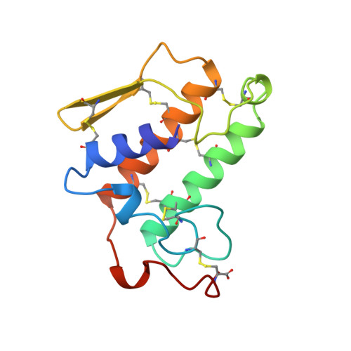
    The trigonal crystal structure of the recombinant bovine pancreatic phospholipase A2 has been re-refined at a slightly higher resolution (1.72 A). The crystals are trigonal, space group P3121, unit-cell parameters a = b = 46.78 and c = 102.89 A and are isomorphous to the previous structure. The structure was refined to a final crystallographic R value of 19.5% (Rfree = 28.4%) using 10 531 reflections. A total of 106 solvent molecules were included in the refinement compared with the earlier refinement which contains only 85 water molecules and 8 925 reflections at 1.8 A resolution. The root-mean-square deviation from the ideal bond lengths and bond angles is considerably better in the present refinement. The active site is extended ( approximately 14 A) from Ala1 to the calcium. The three catalytic residues (Asp99, His48 and the catalytic water) are connected by the conserved structural water and the N-terminal Ala1 on one side, and by the calcium through an equatorial water on the other. The water molecules play a role in the activity of the enzyme PLA2. The Ala1 end of the extended active site performs the activation of the phospholid membranes while the opposite end performs the hydrolysis of the monomeric phospholids.


  • Organizational Affiliation

    Biological Macromolecular Structure Center, Departments of Chemistry and Biochemistry and the Ohio State Biochemistry Program, 100 West 18th Avenue, The Ohio State University, Columbus, OH 43210, USA.


Macromolecules
Find similar proteins by:  (by identity cutoff)  |  3D Structure
Entity ID: 1
MoleculeChains Sequence LengthOrganismDetailsImage
PHOSPHOLIPASE A2123Bos taurusMutation(s): 0 
Gene Names: MATURE PLA2
EC: 3.1.1.4
UniProt
Find proteins for P00593 (Bos taurus)
Explore P00593 
Go to UniProtKB:  P00593
Entity Groups  
Sequence Clusters30% Identity50% Identity70% Identity90% Identity95% Identity100% Identity
UniProt GroupP00593
Sequence Annotations
Expand
  • Reference Sequence
Small Molecules
Ligands 1 Unique
IDChains Name / Formula / InChI Key2D Diagram3D Interactions
CA
Query on CA

Download Ideal Coordinates CCD File 
B [auth A]CALCIUM ION
Ca
BHPQYMZQTOCNFJ-UHFFFAOYSA-N
Experimental Data & Validation

Experimental Data

  • Method: X-RAY DIFFRACTION
  • Resolution: 1.72 Å
  • R-Value Free: 0.284 
  • R-Value Work: 0.195 
  • R-Value Observed: 0.195 
  • Space Group: P 31 2 1
Unit Cell:
Length ( Å )Angle ( ˚ )
a = 46.78α = 90
b = 46.78β = 90
c = 102.89γ = 120
Software Package:
Software NamePurpose
R-AXISdata collection
R-AXISdata reduction
X-PLORmodel building
X-PLORrefinement
R-AXISdata scaling
X-PLORphasing

Structure Validation

View Full Validation Report


Entry History 

Deposition Data

Revision History  (Full details and data files)

  • Version 1.0: 1998-03-11
    Type: Initial release
  • Version 1.1: 2008-03-24
    Changes: Version format compliance
  • Version 1.2: 2011-07-13
    Changes: Version format compliance
  • Version 1.3: 2018-04-04
    Changes: Data collection, Other
  • Version 1.4: 2018-04-11
    Changes: Data collection
  • Version 1.5: 2023-08-09
    Changes: Database references, Derived calculations, Refinement description