The NMR solution conformation of unligated human cyclophilin A.
Ottiger, M., Zerbe, O., Guntert, P., Wuthrich, K.(1997) J Mol Biol 272: 64-81
- PubMed: 9299338
- DOI: https://doi.org/10.1006/jmbi.1997.1220
- PubMed Abstract:
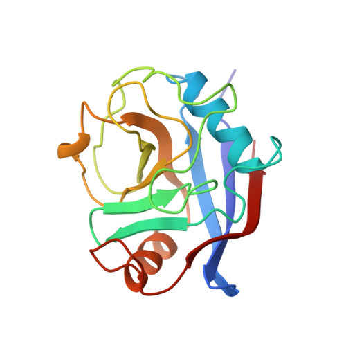
The nuclear magnetic resonance (NMR) solution structure of free, unligated cyclophilin A (CypA), which is an 18 kDa protein from human T-lymphocytes that was expressed in Escherichia coli for the present study, was determined using multidimensional heteronuclear NMR techniques. Sequence-specific resonance assignments for 99.5% of all backbone amide protons and non-labile hydrogen atoms provided the basis for collection of an input of 4101 nuclear Overhauser enhancement (NOE) upper distance constraints and 371 dihedral angle constraints for distance geometry calculations and energy minimization with the programs DIANA and OPAL. The average RMSD values of the 20 best energy-refined NMR conformers relative to the mean coordinates are 0.49 A for the backbone atoms and 0.88 A for all heavy atoms of residues 2 to 165. The molecular architecture includes an eight-stranded antiparallel beta-barrel that is closed by two amphipathic alpha-helices. Detailed comparisons with the crystal structure of free CypA revealed subtle but significant conformational differences that can in most cases be related to lattice contacts in the crystal structure. 15N spin relaxation times and NMR lineshape analyses for CypA in the free form and complexed with cyclosporin A (CsA) revealed transitions of polypeptide loops surrounding the ligand-binding site from locally flexible conformations in the free protein, some of which include well-defined conformational equilibria, to well-defined spatial arrangements in the CypA-CsA complex. Compared to the crystal structure of free CypA, where the ligand-binding area is extensively involved in lattice contacts, the NMR structure presents a highly relevant reference for studies of changes in structure and internal mobility of the binding pocket upon ligand binding, and possible consequences of this conformational variability for calcineurin recognition by the CypA-CsA complex.
Organizational Affiliation:
Institut für Molekularbiologie und Biophysik, Eidgenössische Technische Hochschule Hönggerberg, Zürich, CH-8093, Switzerland.