1PKF

Crystal Structure of Epothilone D-bound Cytochrome P450epoK


Experimental Data Snapshot

  • Method: X-RAY DIFFRACTION
  • Resolution: 2.10 Å
  • R-Value Free: 0.275 
  • R-Value Work: 0.219 
  • R-Value Observed: 0.220 

wwPDB Validation   3D Report Full Report


Ligand Structure Quality Assessment 


This is version 1.4 of the entry. See complete history


Literature

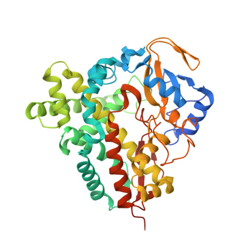
Crystal Structures of Epothilone D-bound, Epothilone B-bound, and Substrate-free Forms of Cytochrome P450epoK

Nagano, S.Li, H.Shimizu, H.Nishida, C.Ogura, H.Ortiz de Montellano, P.R.Poulos, T.L.

(2003) J Biol Chem 278: 44886-44893

  • DOI: https://doi.org/10.1074/jbc.M308115200
  • Primary Citation of Related Structures:  
    1PKF, 1Q5D, 1Q5E

  • PubMed Abstract: 

    Epothilones are potential anticancer drugs that stabilize microtubules by binding to tubulin in a manner similar to paclitaxel. Cytochrome P450epoK (P450epoK), a heme containing monooxygenase involved in epothilone biosynthesis in the myxobacterium Sorangium cellulosum, catalyzes the epoxidation of epothilones C and D into epothilones A and B, respectively. The 2.10-, 1.93-, and 2.65-A crystal structures reported here for the epothilone D-bound, epothilone B-bound, and substrate-free forms, respectively, are the first crystal structures of an epothilone-binding protein. Although the substrate for P450epoK is the largest of a P450 whose x-ray structure is known, the structural changes along with substrate binding or product release are very minor and the overall fold is similar to other P450s. The epothilones are positioned with the macrolide ring roughly perpendicular to the heme plane and I helix, and the thiazole moiety provides key interactions that very likely are critical in determining substrate specificity. Interestingly, there are strong parallels between the epothilone/P450epoK and paclitaxel/tubulin interactions. Based on structural similarities, a plausible epothilone tubulin-binding mode is proposed.


  • Organizational Affiliation

    Department of Molecular Biology & Biochemistry, University of California, Irvine, 92697-3900, USA.


Macromolecules
Find similar proteins by:  (by identity cutoff)  |  3D Structure
Entity ID: 1
MoleculeChains Sequence LengthOrganismDetailsImage
cytochrome p450EpoK419Sorangium cellulosumMutation(s): 0 
UniProt
Find proteins for Q9KIZ4 (Sorangium cellulosum)
Explore Q9KIZ4 
Go to UniProtKB:  Q9KIZ4
Entity Groups  
Sequence Clusters30% Identity50% Identity70% Identity90% Identity95% Identity100% Identity
UniProt GroupQ9KIZ4
Sequence Annotations
Expand
  • Reference Sequence
Small Molecules
Ligands 2 Unique
IDChains Name / Formula / InChI Key2D Diagram3D Interactions
HEM
Query on HEM

Download Ideal Coordinates CCD File 
B [auth A]PROTOPORPHYRIN IX CONTAINING FE
C34 H32 Fe N4 O4
KABFMIBPWCXCRK-RGGAHWMASA-L
EPD
Query on EPD

Download Ideal Coordinates CCD File 
C [auth A]EPOTHILONE D
C27 H41 N O5 S
XOZIUKBZLSUILX-GIQCAXHBSA-N
Experimental Data & Validation

Experimental Data

  • Method: X-RAY DIFFRACTION
  • Resolution: 2.10 Å
  • R-Value Free: 0.275 
  • R-Value Work: 0.219 
  • R-Value Observed: 0.220 
  • Space Group: P 43 2 2
Unit Cell:
Length ( Å )Angle ( ˚ )
a = 60.43α = 90
b = 60.43β = 90
c = 252.77γ = 90
Software Package:
Software NamePurpose
REFMACrefinement
HKL-2000data reduction

Structure Validation

View Full Validation Report


Ligand Structure Quality Assessment 


Entry History 

Deposition Data

Revision History  (Full details and data files)

  • Version 1.0: 2003-10-28
    Type: Initial release
  • Version 1.1: 2008-04-29
    Changes: Version format compliance
  • Version 1.2: 2011-07-13
    Changes: Version format compliance
  • Version 1.3: 2018-01-31
    Changes: Experimental preparation
  • Version 1.4: 2024-02-14
    Changes: Data collection, Database references, Derived calculations