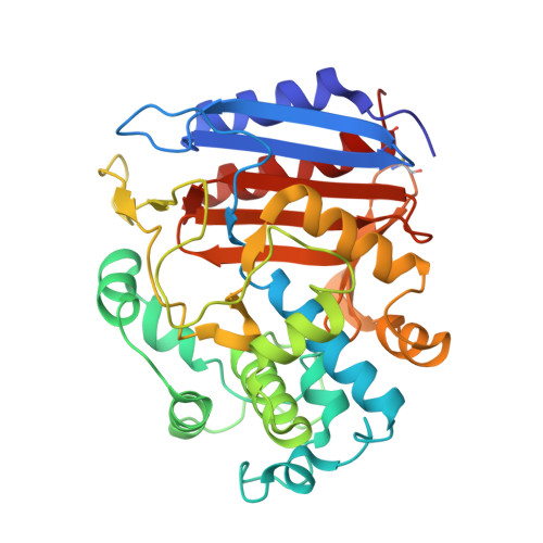
Crystal structures of complexes between the R61 DD-peptidase and peptidoglycan-mimetic beta-lactams: a non-covalent complex with a "perfect penicillin"
Silvaggi, N.R., Josephine, H.R., Kuzin, A.P., Nagarajan, R., Pratt, R.F., Kelly, J.A.(2005) J Mol Biol 345: 521-533
- PubMed: 15581896
- DOI: https://doi.org/10.1016/j.jmb.2004.10.076
- Primary Citation of Related Structures:
1PW1, 1PW8, 1PWC, 1PWD, 1PWG - PubMed Abstract:
The bacterial D-alanyl-D-alanine transpeptidases (DD-peptidases) are the killing targets of beta-lactams, the most important clinical defense against bacterial infections. However, due to the constant development of antibiotic-resistance mechanisms by bacteria, there is an ever-present need for new, more effective antimicrobial drugs. While enormous numbers of beta-lactam compounds have been tested for antibiotic activity in over 50 years of research, the success of a beta-lactam structure in terms of antibiotic activity remains unpredictable. Tipper and Strominger suggested long ago that beta-lactams inhibit DD-peptidases because they mimic the D-alanyl-D-alanine motif of the peptidoglycan substrate of these enzymes. They also predicted that beta-lactams having a peptidoglycan-mimetic side-chain might be better antibiotics than their non-specific counterparts, but decades of research have not provided any evidence for this. We have recently described two such novel beta-lactams. The first is a penicillin having the glycyl-L-alpha-amino-epsilon-pimelyl side-chain of Streptomyces strain R61 peptidoglycan, making it the "perfect penicillin" for this organism. The other is a cephalosporin with the same side-chain. Here, we describe the X-ray crystal structures of the perfect penicillin in non-covalent and covalent complexes with the Streptomyces R61 DD-peptidase. The structure of the non-covalent enzyme-inhibitor complex is the first such complex to be trapped crystallographically with a DD-peptidase. In addition, the covalent complex of the peptidyl-cephalosporin with the R61 DD-peptidase is described. Finally, two covalent complexes with the traditional beta-lactams benzylpenicillin and cephalosporin C were determined for comparison with the peptidyl beta-lactams. These structures, together with relevant kinetics data, support Tipper and Strominger's assertion that peptidoglycan-mimetic side-chains should improve beta-lactams as inhibitors of DD-peptidases.
Organizational Affiliation:
Department of Molecular and Cell Biology and Institute for Materials Science, University of Connecticut, Storrs, CT 06269-3125, USA.