1Q13

Crystal structure of rabbit 20alpha hyroxysteroid dehydrogenase in ternary complex with NADP and testosterone


Experimental Data Snapshot

  • Method: X-RAY DIFFRACTION
  • Resolution: 2.08 Å
  • R-Value Free: 0.280 
  • R-Value Work: 0.226 
  • R-Value Observed: 0.229 

wwPDB Validation   3D Report Full Report


Ligand Structure Quality Assessment 


This is version 1.5 of the entry. See complete history


Literature

Loop relaxation, a mechanism that explains the reduced specificity of rabbit 20alpha-hydroxysteroid dehydrogenase, a member of the aldo-keto reductase superfamily.

Couture, J.F.Legrand, P.Cantin, L.Labrie, F.Luu-The, V.Breton, R.

(2004) J Mol Biol 339: 89-102

  • DOI: https://doi.org/10.1016/j.jmb.2004.03.035
  • Primary Citation of Related Structures:  
    1Q13, 1Q5M

  • PubMed Abstract: 

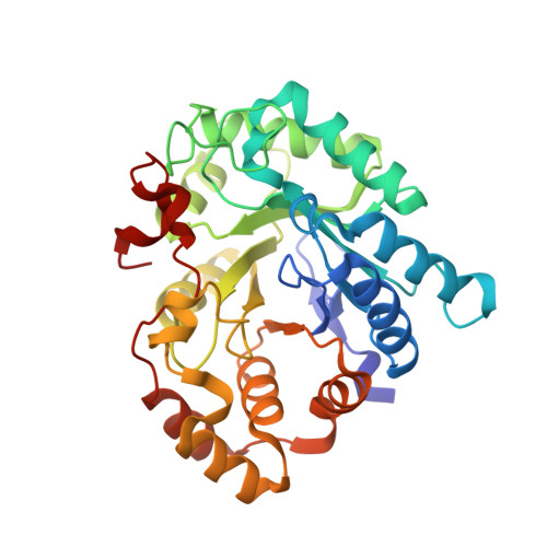
    The aldo-keto reductase rabbit 20alpha-hydroxysteroid dehydrogenase (rb20alpha-HSD; AKR1C5) is less selective than other HSDs, since it exerts its activity both on androgens (C19 steroids) and progestins (C21 steroids). In order to identify the molecular determinants responsible for this reduced selectivity, binary (NADPH) and ternary (NADP(+)/testosterone) complex structures were solved to 1.32A and 2.08A resolution, respectively. Inspection of the cofactor-binding cavity led to the identification of a new interaction between side-chains of residues His222 and Lys270, which cover the central phosphate chain of the cofactor, reminiscent of the "safety-belt" found in other aldo-keto reductases. Testosterone is stabilized by a phenol/benzene tunnel composed of side-chains of numerous residues, among which Phe54, which forces the steroid to take up an orientation markedly contrasting with that found in HSD ternary complexes reported. Combining structural, site-directed mutagenesis, kinetic and fluorescence titration studies, we found that the selectivity of rb20alpha-HSD is mediated by (i) the relaxation of loop B (residues 223-230), partly controlled by the nature of residue 230, (ii) the nature of the residue found at position 54, and (iii) the residues found in the C-terminal tail of the protein especially the side-chain of the amino acid 306.


  • Organizational Affiliation

    Oncology and Molecular Endocrinology Research Center, Laval University Medical Center (CHUL) and Laval University, Ste-Foy, Quebec, Canada G1V 4G2.


Macromolecules
Find similar proteins by:  (by identity cutoff)  |  3D Structure
Entity ID: 1
MoleculeChains Sequence LengthOrganismDetailsImage
Prostaglandin-E2 9-reductase
A, B
323Oryctolagus cuniculusMutation(s): 0 
EC: 1.1.1.189 (PDB Primary Data), 1.1.1.149 (PDB Primary Data)
UniProt
Find proteins for P80508 (Oryctolagus cuniculus)
Explore P80508 
Go to UniProtKB:  P80508
Entity Groups  
Sequence Clusters30% Identity50% Identity70% Identity90% Identity95% Identity100% Identity
UniProt GroupP80508
Sequence Annotations
Expand
  • Reference Sequence
Experimental Data & Validation

Experimental Data

  • Method: X-RAY DIFFRACTION
  • Resolution: 2.08 Å
  • R-Value Free: 0.280 
  • R-Value Work: 0.226 
  • R-Value Observed: 0.229 
  • Space Group: P 1 21 1
Unit Cell:
Length ( Å )Angle ( ˚ )
a = 57.21α = 90
b = 84.046β = 91.17
c = 66.249γ = 90
Software Package:
Software NamePurpose
REFMACrefinement
XDSdata scaling
AMoREphasing

Structure Validation

View Full Validation Report


Ligand Structure Quality Assessment 


Entry History 

Deposition Data

Revision History  (Full details and data files)

  • Version 1.0: 2004-11-16
    Type: Initial release
  • Version 1.1: 2008-04-29
    Changes: Version format compliance
  • Version 1.2: 2011-07-13
    Changes: Advisory, Version format compliance
  • Version 1.3: 2017-10-11
    Changes: Refinement description
  • Version 1.4: 2017-12-20
    Changes: Database references
  • Version 1.5: 2024-02-14
    Changes: Data collection, Database references, Derived calculations, Refinement description