1Q7A

Crystal structure of the complex formed between russell's viper phospholipase A2 and an antiinflammatory agent oxyphenbutazone at 1.6A resolution


Experimental Data Snapshot

  • Method: X-RAY DIFFRACTION
  • Resolution: 1.60 Å
  • R-Value Free: 0.213 
  • R-Value Work: 0.209 
  • R-Value Observed: 0.209 

wwPDB Validation   3D Report Full Report


This is version 1.3 of the entry. See complete history


Literature

Phospholipase A2 as a target protein for nonsteroidal anti-inflammatory drugs (NSAIDS): crystal structure of the complex formed between phospholipase A2 and oxyphenbutazone at 1.6 A resolution.

Singh, N.Jabeen, T.Somvanshi, R.K.Sharma, S.Dey, S.Singh, T.P.

(2004) Biochemistry 43: 14577-14583

  • DOI: https://doi.org/10.1021/bi0483561
  • Primary Citation of Related Structures:  
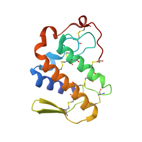
    1Q7A

  • PubMed Abstract: 

    Phospholipase A(2) (PLA(2); EC 3.1.1.4) is a key enzyme involved in the production of proinflammatory mediators known as eicosanoids. The binding of the substrate to PLA(2) occurs through a well-formed hydrophobic channel. To determine the viability of PLA(2) as a target molecule for the structure-based drug design against inflammation, arthritis, and rheumatism, the crystal structure of the complex of PLA(2) with a known anti-inflammatory compound oxyphenbutazone (OPB), which has been determined at 1.6 A resolution. The structure has been refined to an R factor of 0.209. The structure contains 1 molecule each of PLA(2) and OPB with 2 sulfate ions and 111 water molecules. The binding studies using surface plasmon resonance show that OPB binds to PLA(2) with a dissociation constant of 6.4 x 10(-8) M. The structure determination has revealed the presence of an OPB molecule at the binding site of PLA(2). It fits well in the binding region, thus displaying a high level of complementarity. The structure also indicates that OPB works as a competitive inhibitor. A large number of hydrophobic interactions between the enzyme and the OPB molecule have been observed. The hydrophobic interactions involving residues Tyr(52) and Lys(69) with OPB are particularly noteworthy. Other residues of the hydrophobic channel such as Leu(3), Phe(5), Met(8), Ile(9), and Ala(18) are also interacting extensively with the inhibitor. The crystal structure clearly reveals that the binding of OPB to PLA(2) is specific in nature and possibly suggests that the basis of its anti-inflammatory effects may be due to its binding to PLA(2) as well.


  • Organizational Affiliation

    Department of Biophysics, All India Institute of Medical Sciences, New Delhi 110029, India.


Macromolecules
Find similar proteins by:  (by identity cutoff)  |  3D Structure
Entity ID: 1
MoleculeChains Sequence LengthOrganismDetailsImage
Phospholipase A2 VRV-PL-VIIIa121Daboia russelii pulchellaMutation(s): 0 
EC: 3.1.1.4
UniProt
Find proteins for P59071 (Daboia russelii)
Explore P59071 
Go to UniProtKB:  P59071
Entity Groups  
Sequence Clusters30% Identity50% Identity70% Identity90% Identity95% Identity100% Identity
UniProt GroupP59071
Sequence Annotations
Expand
  • Reference Sequence
Small Molecules
Ligands 3 Unique
IDChains Name / Formula / InChI Key2D Diagram3D Interactions
OPB
Query on OPB

Download Ideal Coordinates CCD File 
D [auth A]4-BUTYL-1-(4-HYDROXYPHENYL)-2-PHENYLPYRAZOLIDINE-3,5-DIONE
C19 H20 N2 O3
HFHZKZSRXITVMK-KRWDZBQOSA-N
SO4
Query on SO4

Download Ideal Coordinates CCD File 
B [auth A],
C [auth A]
SULFATE ION
O4 S
QAOWNCQODCNURD-UHFFFAOYSA-L
MOH
Query on MOH

Download Ideal Coordinates CCD File 
E [auth A]METHANOL
C H4 O
OKKJLVBELUTLKV-UHFFFAOYSA-N
Binding Affinity Annotations 
IDSourceBinding Affinity
OPB PDBBind:  1Q7A Kd: 64 (nM) from 1 assay(s)
Binding MOAD:  1Q7A Kd: 64 (nM) from 1 assay(s)
Experimental Data & Validation

Experimental Data

  • Method: X-RAY DIFFRACTION
  • Resolution: 1.60 Å
  • R-Value Free: 0.213 
  • R-Value Work: 0.209 
  • R-Value Observed: 0.209 
  • Space Group: P 43
Unit Cell:
Length ( Å )Angle ( ˚ )
a = 53α = 90
b = 53β = 90
c = 48.2γ = 90
Software Package:
Software NamePurpose
CNSrefinement
DENZOdata reduction
SCALEPACKdata scaling
AMoREphasing

Structure Validation

View Full Validation Report


Entry History 

Deposition Data

Revision History  (Full details and data files)

  • Version 1.0: 2004-05-11
    Type: Initial release
  • Version 1.1: 2008-04-29
    Changes: Version format compliance
  • Version 1.2: 2011-07-13
    Changes: Version format compliance
  • Version 1.3: 2023-08-16
    Changes: Data collection, Database references, Derived calculations, Refinement description