1QW4

Crystal Structure of Murine Inducible Nitric Oxide Synthase Oxygenase Domain in complex with N-omega-propyl-L-arginine.


Experimental Data Snapshot

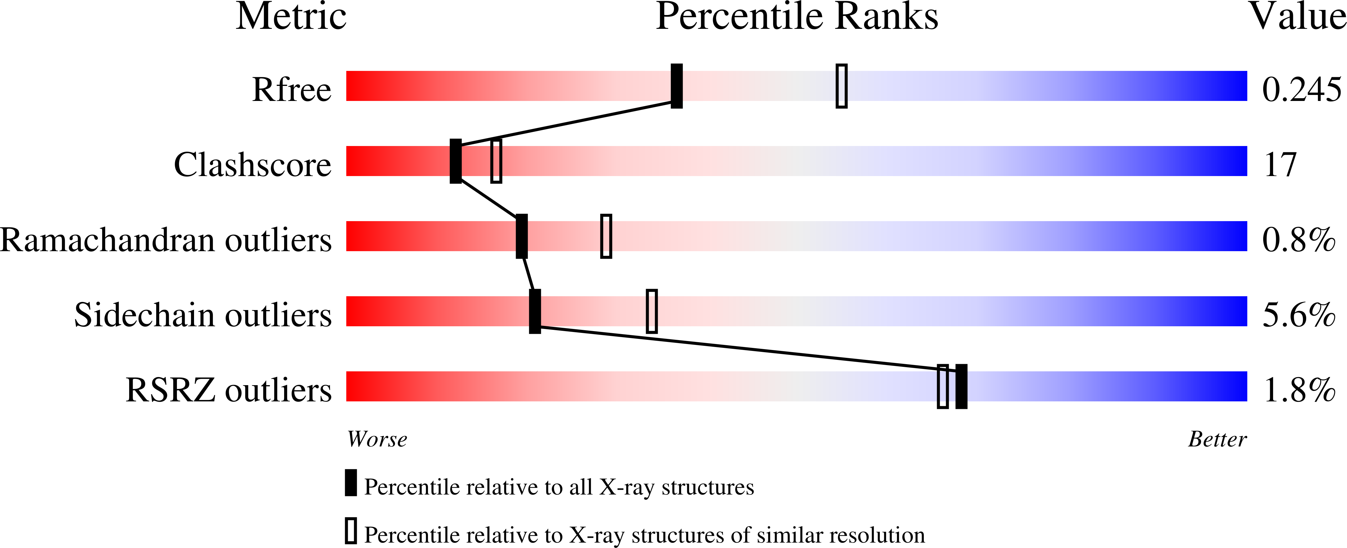
  • Method: X-RAY DIFFRACTION
  • Resolution: 2.40 Å
  • R-Value Free: 0.251 
  • R-Value Work: 0.213 
  • R-Value Observed: 0.215 

wwPDB Validation   3D Report Full Report


Ligand Structure Quality Assessment 


This is version 1.4 of the entry. See complete history


Literature

Structural basis for the specificity of the nitric-oxide synthase inhibitors W1400 and Nomega-propyl-L-Arg for the inducible and neuronal isoforms.

Fedorov, R.Hartmann, E.Ghosh, D.K.Schlichting, I.

(2003) J Biol Chem 278: 45818-45825

  • DOI: https://doi.org/10.1074/jbc.M306030200
  • Primary Citation of Related Structures:  
    1QW4, 1QW5, 1QW6, 1QWC

  • PubMed Abstract: 

    The high level of amino acid conservation and structural similarity in the immediate vicinity of the substrate binding sites of the oxygenase domains of the nitric-oxide synthase (NOS) isoforms (eNOSoxy, iNOSoxy, and nNOSoxy) make the interpretation of the structural basis of inhibitor isoform specificity a challenge and provide few clues for the design of new selective compounds. Crystal structures of iNOSoxy and nNOSoxy complexed with the inhibitors W1400 and Nomega-propyl-l-arginine provide a rationale for their isoform specificity. It involves differences outside the immediate active site as well as a conformational flexibility in the active site that allows the adoption of distinct conformations in response to interactions with the inhibitors. This flexibility is determined by isoform-specific residues outside the active site.


  • Organizational Affiliation

    Max Planck Institut für Molekulare Physiologie, Abteilung Biophysikalische Chemie, Otto Hahn Strasse 11, 44227 Dortmund, Germany.


Macromolecules
Find similar proteins by:  (by identity cutoff)  |  3D Structure
Entity ID: 1
MoleculeChains Sequence LengthOrganismDetailsImage
Nitric oxide synthase, inducible
A, B
419Mus musculusMutation(s): 0 
Gene Names: NOS2 OR INOSL
EC: 1.14.13.39
UniProt & NIH Common Fund Data Resources
Find proteins for P29477 (Mus musculus)
Explore P29477 
Go to UniProtKB:  P29477
IMPC:  MGI:97361
Entity Groups  
Sequence Clusters30% Identity50% Identity70% Identity90% Identity95% Identity100% Identity
UniProt GroupP29477
Sequence Annotations
Expand
  • Reference Sequence
Small Molecules
Ligands 4 Unique
IDChains Name / Formula / InChI Key2D Diagram3D Interactions
HEM
Query on HEM

Download Ideal Coordinates CCD File 
D [auth A],
H [auth B]
PROTOPORPHYRIN IX CONTAINING FE
C34 H32 Fe N4 O4
KABFMIBPWCXCRK-RGGAHWMASA-L
H4B
Query on H4B

Download Ideal Coordinates CCD File 
E [auth A],
I [auth B]
5,6,7,8-TETRAHYDROBIOPTERIN
C9 H15 N5 O3
FNKQXYHWGSIFBK-RPDRRWSUSA-N
3AR
Query on 3AR

Download Ideal Coordinates CCD File 
F [auth A],
J [auth B]
N-OMEGA-PROPYL-L-ARGININE
C9 H21 N4 O2
AOMXURITGZJPKB-ZETCQYMHSA-O
ZN
Query on ZN

Download Ideal Coordinates CCD File 
C [auth A],
G [auth B]
ZINC ION
Zn
PTFCDOFLOPIGGS-UHFFFAOYSA-N
Experimental Data & Validation

Experimental Data

  • Method: X-RAY DIFFRACTION
  • Resolution: 2.40 Å
  • R-Value Free: 0.251 
  • R-Value Work: 0.213 
  • R-Value Observed: 0.215 
  • Space Group: P 61 2 2
Unit Cell:
Length ( Å )Angle ( ˚ )
a = 214.603α = 90
b = 214.603β = 90
c = 117.464γ = 120
Software Package:
Software NamePurpose
ProDCdata collection
XDSdata reduction
CNSrefinement
XDSdata scaling
CNSphasing

Structure Validation

View Full Validation Report


Ligand Structure Quality Assessment 


Entry History 

Deposition Data

Revision History  (Full details and data files)

  • Version 1.0: 2003-12-09
    Type: Initial release
  • Version 1.1: 2008-04-29
    Changes: Version format compliance
  • Version 1.2: 2011-07-13
    Changes: Derived calculations, Version format compliance
  • Version 1.3: 2017-10-11
    Changes: Refinement description
  • Version 1.4: 2024-02-14
    Changes: Data collection, Database references, Derived calculations