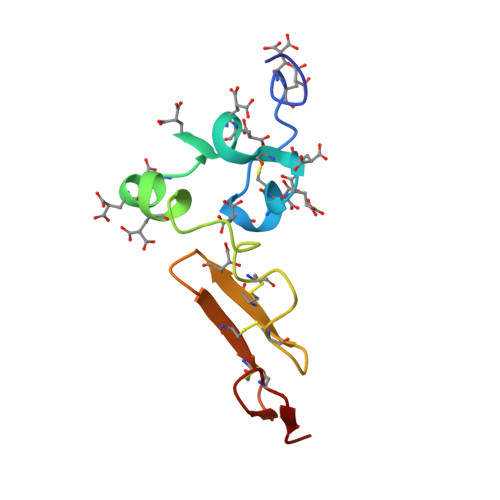
The relative orientation of Gla and EGF domains in coagulation factor X is altered by Ca2+ binding to the first EGF domain. A combined NMR-small angle X-ray scattering study.
Sunnerhagen, M., Olah, G.A., Stenflo, J., Forsen, S., Drakenberg, T., Trewhella, J.(1996) Biochemistry 35: 11547-11559
- PubMed: 8794734
- DOI: https://doi.org/10.1021/bi960633j
- Primary Citation of Related Structures:
1WHE, 1WHF - PubMed Abstract:
Coagulation factor X is a serine protease containing three noncatalytic domains: an N-terminal gamma-carboxyglutamic acid (Gla)1 domain followed by two epidermal growth factor (EGF)-like domains. The isolated N-terminal EGF domain binds Ca2+ with a Kd of 10(-3) M. When linked to the Gla domain, however, its Ca2+ affinity is increased 10-fold. In this paper, we present the NMR solution structure of the factor X Gla-EGF domain pair with Ca2+ bound to the EGF domain, as well as small angle X-ray scattering (SAXS) data on the Gla-EGF domain pair with and without Ca2+. Our results show that Ca2+ binding to the EGF domain makes the Gla and EGF domains fold toward each other using the Ca2+ site as a hinge. Presumably, a similar mechanism may be responsible for alterations in the relative orientation of protein domains in many other extracellular proteins containing EGF domains with the consensus for Ca2+ binding. The results of the NMR and SAXS measurements reported in this paper confirm our previous result that the Gla domain is folded also in its apo state when linked to the EGF domain [Sunnerhagen, M., et al. (1995) Nat. Struct. Biol. 2, 504-509]. Finally, our study clearly demonstrates the powerful combination of NMR and SAXS in the study of modular proteins, since this enables reliable evaluation of both short-range (NMR) and long-range interactions (SAXS).
Organizational Affiliation:
Chemical Center, Lund University, Sweden.


