1XQD

Crystal structure of P450NOR complexed with 3-pyridinealdehyde adenine dinucleotide


Experimental Data Snapshot

  • Method: X-RAY DIFFRACTION
  • Resolution: 1.80 Å
  • R-Value Free: 0.243 
  • R-Value Work: 0.206 
  • R-Value Observed: 0.206 

wwPDB Validation   3D Report Full Report


Ligand Structure Quality Assessment 


This is version 1.4 of the entry. See complete history


Literature

Structural evidence for direct hydride transfer from NADH to cytochrome P450nor

Oshima, R.Fushinobu, S.Su, F.Zhang, L.Takaya, N.Shoun, H.

(2004) J Mol Biol 342: 207-217

  • DOI: https://doi.org/10.1016/j.jmb.2004.07.009
  • Primary Citation of Related Structures:  
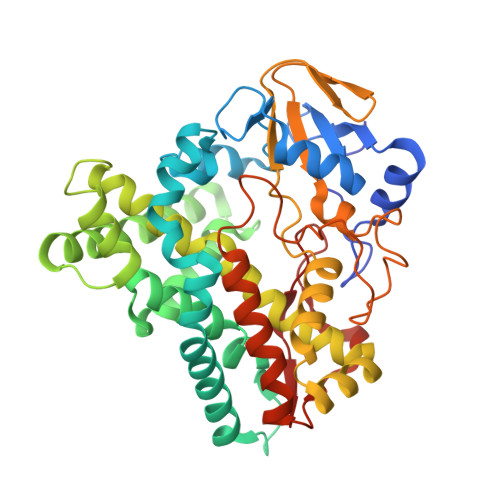
    1ULW, 1XQD

  • PubMed Abstract: 

    Nitric oxide reductase cytochrome P450nor catalyzes an unusual reaction, direct electron transfer from NAD(P)H to bound heme. Here, we succeeded in determining the crystal structure of P450nor in a complex with an NADH analogue, nicotinic acid adenine dinucleotide, which provides conclusive evidence for the mechanism of the unprecedented electron transfer. Comparison of the structure with those of dinucleotide-free forms revealed a global conformational change accompanied by intriguing local movements caused by the binding of the pyridine nucleotide. Arg64 and Arg174 fix the pyrophosphate moiety upon the dinucleotide binding. Stereo-selective hydride transfer from NADH to NO-bound heme was suggested from the structure, the nicotinic acid ring being fixed near the heme by the conserved Thr residue in the I-helix and the upward-shifted propionate side-chain of the heme. A proton channel near the NADH channel is formed upon the dinucleotide binding, which should direct continuous transfer of the hydride and proton. A salt-bridge network (Glu71-Arg64-Asp88) was shown to be crucial for a high catalytic turnover.


  • Organizational Affiliation

    Department of Biotechnology, Graduate School of Agricultural and Life Sciences, The University of Tokyo, 1-1-1 Yayoi, Bunkyo-ku, 113-8657, Japan.


Macromolecules
Find similar proteins by:  (by identity cutoff)  |  3D Structure
Entity ID: 1
MoleculeChains Sequence LengthOrganismDetailsImage
CYTOCHROME P450 55A1403Fusarium oxysporumMutation(s): 2 
EC: 1.7.99.7
UniProt
Find proteins for P23295 (Fusarium oxysporum)
Explore P23295 
Go to UniProtKB:  P23295
Entity Groups  
Sequence Clusters30% Identity50% Identity70% Identity90% Identity95% Identity100% Identity
UniProt GroupP23295
Sequence Annotations
Expand
  • Reference Sequence
Small Molecules
Ligands 2 Unique
IDChains Name / Formula / InChI Key2D Diagram3D Interactions
DND
Query on DND

Download Ideal Coordinates CCD File 
C [auth A]NICOTINIC ACID ADENINE DINUCLEOTIDE
C21 H27 N6 O15 P2
SENPVEZBRZQVST-HISDBWNOSA-O
HEM
Query on HEM

Download Ideal Coordinates CCD File 
B [auth A]PROTOPORPHYRIN IX CONTAINING FE
C34 H32 Fe N4 O4
KABFMIBPWCXCRK-RGGAHWMASA-L
Binding Affinity Annotations 
IDSourceBinding Affinity
DND Binding MOAD:  1XQD Kd: 7.00e+4 (nM) from 1 assay(s)
Experimental Data & Validation

Experimental Data

  • Method: X-RAY DIFFRACTION
  • Resolution: 1.80 Å
  • R-Value Free: 0.243 
  • R-Value Work: 0.206 
  • R-Value Observed: 0.206 
  • Space Group: P 21 21 21
Unit Cell:
Length ( Å )Angle ( ˚ )
a = 43.296α = 90
b = 56.121β = 90
c = 163.262γ = 90
Software Package:
Software NamePurpose
HKL-2000data collection
SCALEPACKdata scaling
MOLREPphasing
CNSrefinement
HKL-2000data reduction

Structure Validation

View Full Validation Report


Ligand Structure Quality Assessment 


Entry History 

Deposition Data

Revision History  (Full details and data files)

  • Version 1.0: 2004-10-26
    Type: Initial release
  • Version 1.1: 2008-04-30
    Changes: Version format compliance
  • Version 1.2: 2011-07-13
    Changes: Version format compliance
  • Version 1.3: 2021-11-10
    Changes: Database references, Derived calculations
  • Version 1.4: 2023-10-25
    Changes: Data collection, Refinement description