2BAX

Atomic Resolution Structure of the Double Mutant (K53,56M) of Bovine Pancreatic Phospholipase A2


Experimental Data Snapshot

  • Method: X-RAY DIFFRACTION
  • Resolution: 1.10 Å
  • R-Value Free: 0.156 
  • R-Value Work: 0.119 
  • R-Value Observed: 0.114 

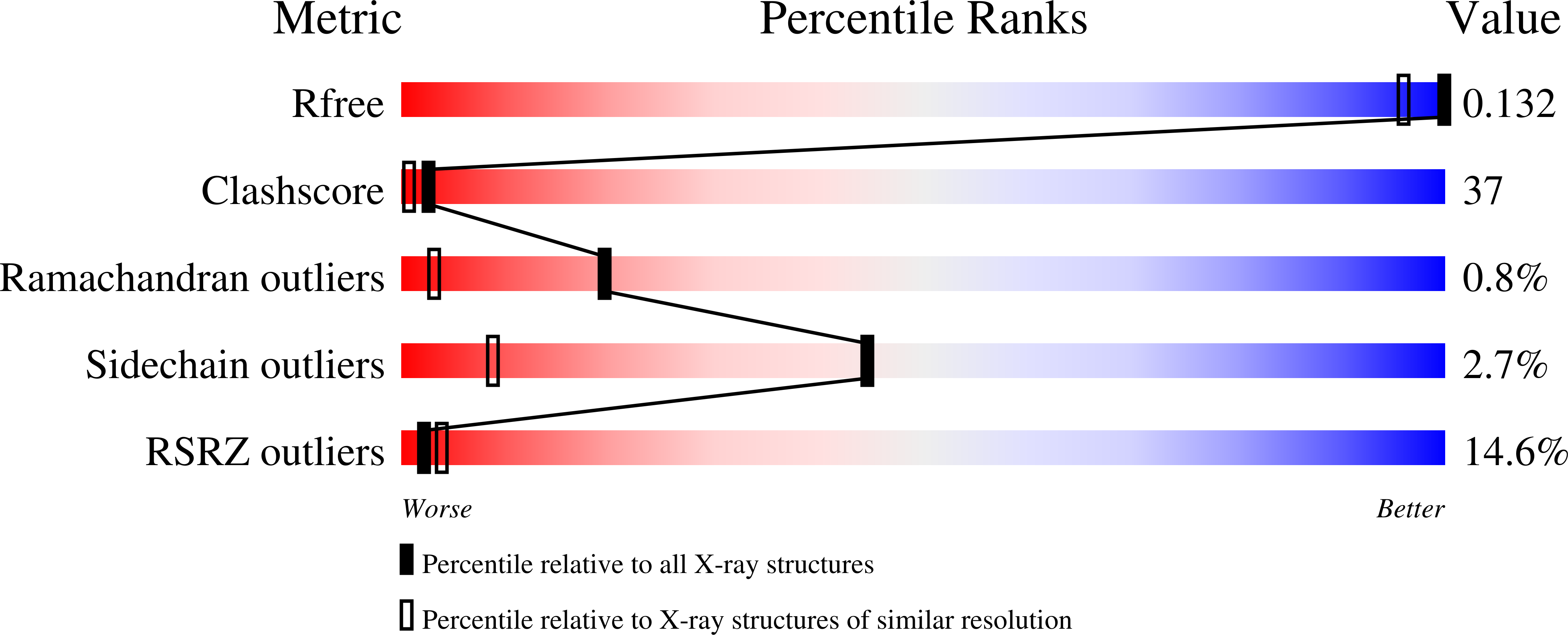
wwPDB Validation   3D Report Full Report


This is version 1.4 of the entry. See complete history


Literature

Atomic resolution (0.97 A) structure of the triple mutant (K53,56,121M) of bovine pancreatic phospholipase A2.

Sekar, K.Rajakannan, V.Gayathri, D.Velmurugan, D.Poi, M.J.Dauter, M.Dauter, Z.Tsai, M.D.

(2005) Acta Crystallogr Sect F Struct Biol Cryst Commun 61: 3-7

  • DOI: https://doi.org/10.1107/S1744309104021748
  • Primary Citation of Related Structures:  
    1VL9, 2BAX

  • PubMed Abstract: 

    The enzyme phospholipase A2 catalyzes the hydrolysis of the sn-2 acyl chain of phospholipids, forming fatty acids and lysophospholipids. The crystal structure of a triple mutant (K53,56,121M) of bovine pancreatic phospholipase A2 in which the lysine residues at positions 53, 56 and 121 are replaced recombinantly by methionines has been determined at atomic resolution (0.97 A). The crystal is monoclinic (space group P2), with unit-cell parameters a = 36.934, b = 23.863, c = 65.931 A, beta = 101.47 degrees. The structure was solved by molecular replacement and has been refined to a final R factor of 10.6% (Rfree = 13.4%) using 63,926 unique reflections. The final protein model consists of 123 amino-acid residues, two calcium ions, one chloride ion, 243 water molecules and six 2-methyl-2,4-pentanediol molecules. The surface-loop residues 60-70 are ordered and have clear electron density.


  • Organizational Affiliation

    Bioinformatics Centre, Indian Institute of Science, Bangalore 560 012, India. sekar@physics.iisc.ernet.in


Macromolecules
Find similar proteins by:  (by identity cutoff)  |  3D Structure
Entity ID: 1
MoleculeChains Sequence LengthOrganismDetailsImage
Phospholipase A2123Bos taurusMutation(s): 2 
Gene Names: PLA2G1B
EC: 3.1.1.4
UniProt
Find proteins for P00593 (Bos taurus)
Explore P00593 
Go to UniProtKB:  P00593
Entity Groups  
Sequence Clusters30% Identity50% Identity70% Identity90% Identity95% Identity100% Identity
UniProt GroupP00593
Sequence Annotations
Expand
  • Reference Sequence
Small Molecules
Ligands 4 Unique
IDChains Name / Formula / InChI Key2D Diagram3D Interactions
MPD
Query on MPD

Download Ideal Coordinates CCD File 
H [auth A](4S)-2-METHYL-2,4-PENTANEDIOL
C6 H14 O2
SVTBMSDMJJWYQN-YFKPBYRVSA-N
MRD
Query on MRD

Download Ideal Coordinates CCD File 
D [auth A],
E [auth A],
F [auth A],
G [auth A]
(4R)-2-METHYLPENTANE-2,4-DIOL
C6 H14 O2
SVTBMSDMJJWYQN-RXMQYKEDSA-N
CA
Query on CA

Download Ideal Coordinates CCD File 
B [auth A]CALCIUM ION
Ca
BHPQYMZQTOCNFJ-UHFFFAOYSA-N
CL
Query on CL

Download Ideal Coordinates CCD File 
C [auth A]CHLORIDE ION
Cl
VEXZGXHMUGYJMC-UHFFFAOYSA-M
Experimental Data & Validation

Experimental Data

  • Method: X-RAY DIFFRACTION
  • Resolution: 1.10 Å
  • R-Value Free: 0.156 
  • R-Value Work: 0.119 
  • R-Value Observed: 0.114 
  • Space Group: P 31 2 1
Unit Cell:
Length ( Å )Angle ( ˚ )
a = 46.057α = 90
b = 46.057β = 90
c = 101.138γ = 120
Software Package:
Software NamePurpose
HKL-2000data collection
SCALAdata scaling
MOLREPphasing
REFMACrefinement
HKL-2000data reduction
CCP4data scaling

Structure Validation

View Full Validation Report


Entry History 

Deposition Data

Revision History  (Full details and data files)

  • Version 1.0: 2005-10-25
    Type: Initial release
  • Version 1.1: 2008-05-01
    Changes: Version format compliance
  • Version 1.2: 2011-07-13
    Changes: Version format compliance
  • Version 1.3: 2021-10-20
    Changes: Database references, Derived calculations
  • Version 1.4: 2023-08-23
    Changes: Data collection, Refinement description