2CYP

Crystal structure of yeast cytochrome C peroxidase refined at 1.7-angstroms resolution


Experimental Data Snapshot

  • Method: X-RAY DIFFRACTION
  • Resolution: 1.70 Å
  • R-Value Work: 0.202 

wwPDB Validation   3D Report Full Report


Ligand Structure Quality Assessment 


This is version 1.4 of the entry. See complete history


Literature

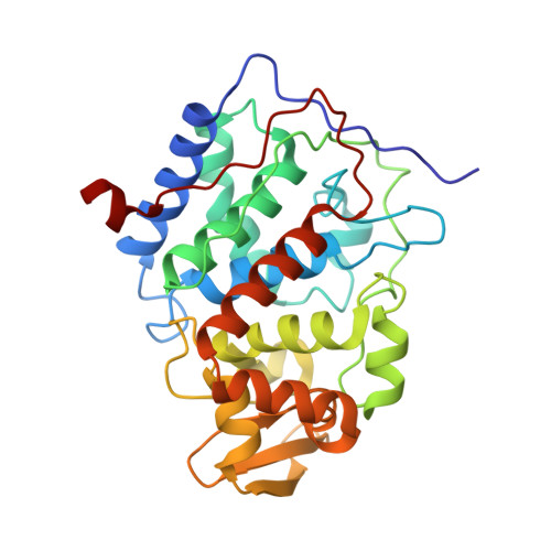
Crystal structure of yeast cytochrome c peroxidase refined at 1.7-A resolution

Finzel, B.C.Poulos, T.L.Kraut, J.

(1984) J Biol Chem 259: 13027-13036

  • Primary Citation of Related Structures:  
    2CYP

  • PubMed Abstract: 

    The crystal structure of cytochrome c peroxidase (EC 1.11.1.5) has been refined to an R factor of 0.20 computed for all reflections to 1.7 A. The refined molecular model includes 263 bound water molecules and allows for x-ray scattering by amorphous solvent. The mean positional error in atomic coordinates is estimated to lie between 0.12 and 0.21 A. Two factors are identified which may account for the ability of the enzyme to stabilize high-oxidation states of the heme iron during catalysis: 1) the proximal histidine forms a hydrogen bond with a buried aspartic acid side chain, Asp-235; and 2) the heme environment is more polar than in the cytochromes c or globins, owing to the presence of the partially buried side-chain of Arg-48 and five water molecules bound in close proximity to the heme. Two of these occupy the presumed peroxide-binding site. Two candidates are likely for the side chain that is oxidized to a free radical during formation of Compound I: 1) Trp-51, which rests 3.3 A above the heme plane in close proximity (2.7 A) to the sixth coordination position; and 2) Met-172, which is 3.7 A from the heme. Nucleophilic stabilization of the methionyl cation radical may be possible via Asp-235. His-181 is found to lie coplanar with the heme in a niche between the two propionates near the suspected cytochrome c-binding site. A network of hydrogen bonds involving this histidine may provide a preferred pathway for electron transfer between hemes.


Macromolecules
Find similar proteins by:  (by identity cutoff)  |  3D Structure
Entity ID: 1
MoleculeChains Sequence LengthOrganismDetailsImage
CYTOCHROME C PEROXIDASE294Saccharomyces cerevisiaeMutation(s): 0 
EC: 1.11.1.5
UniProt
Find proteins for P00431 (Saccharomyces cerevisiae (strain ATCC 204508 / S288c))
Explore P00431 
Go to UniProtKB:  P00431
Entity Groups  
Sequence Clusters30% Identity50% Identity70% Identity90% Identity95% Identity100% Identity
UniProt GroupP00431
Sequence Annotations
Expand
  • Reference Sequence
Small Molecules
Ligands 2 Unique
IDChains Name / Formula / InChI Key2D Diagram3D Interactions
HEM
Query on HEM

Download Ideal Coordinates CCD File 
B [auth A]PROTOPORPHYRIN IX CONTAINING FE
C34 H32 Fe N4 O4
KABFMIBPWCXCRK-RGGAHWMASA-L
O
Query on O

Download Ideal Coordinates CCD File 
C [auth A]OXYGEN ATOM
O
XLYOFNOQVPJJNP-UHFFFAOYSA-N
Experimental Data & Validation

Experimental Data

  • Method: X-RAY DIFFRACTION
  • Resolution: 1.70 Å
  • R-Value Work: 0.202 
  • Space Group: P 21 21 21
Unit Cell:
Length ( Å )Angle ( ˚ )
a = 107.4α = 90
b = 76.8β = 90
c = 51.4γ = 90
Software Package:
Software NamePurpose
PROLSQrefinement

Structure Validation

View Full Validation Report


Ligand Structure Quality Assessment 


Entry History 

Deposition Data

Revision History  (Full details and data files)

  • Version 1.0: 1986-01-21
    Type: Initial release
  • Version 1.1: 2008-03-10
    Changes: Version format compliance
  • Version 1.2: 2011-07-13
    Changes: Version format compliance
  • Version 1.3: 2017-11-29
    Changes: Derived calculations, Other
  • Version 1.4: 2024-02-14
    Changes: Data collection, Database references, Derived calculations