2FSZ

A second binding site for hydroxytamoxifen within the coactivator-binding groove of estrogen receptor beta


Experimental Data Snapshot

  • Method: X-RAY DIFFRACTION
  • Resolution: 2.20 Å
  • R-Value Free: 0.299 
  • R-Value Work: 0.273 
  • R-Value Observed: 0.274 

wwPDB Validation   3D Report Full Report


This is version 1.5 of the entry. See complete history


Literature

A second binding site for hydroxytamoxifen within the coactivator-binding groove of estrogen receptor beta

Wang, Y.Chirgadze, N.Y.Briggs, S.L.Khan, S.Jensen, E.V.Burris, T.P.

(2006) Proc Natl Acad Sci U S A 103: 9908-9911

  • DOI: https://doi.org/10.1073/pnas.0510596103
  • Primary Citation of Related Structures:  
    2FSZ

  • PubMed Abstract: 

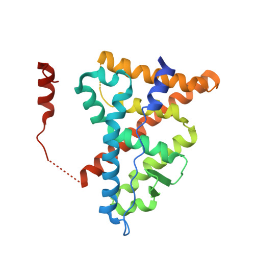
    Evidence is presented that the estrogen antagonist 4-hydroxytamoxifen (HT) can occupy not only the core binding pocket within the ligand-binding domain of estrogen receptor (ER) beta but also a second site on its surface. The crystal structure of the ligand-binding domain (LBD) associated with HT was determined to 2.2 A and revealed two molecules of HT bound to the protein. One was located in the consensus ligand-binding pocket, whereas the other bound to a site that overlaps with the hydrophobic groove of the coactivator recognition surface. Relative to the ERalpha-tamoxifen structure, helix 12 has been displaced from the coactivator recognition surface and occupies a unique position. Although it has been demonstrated that association of the antagonist with the core ligand-binding pocket is sufficient to induce an antagonist ligand-binding domain conformation, this structure suggests that small molecules may directly antagonize receptor-coactivator interactions. These results provide a direct demonstration of two binding sites for HT in ERbeta, as has been previously suggested for ERalpha by using biochemical methods, and represent a crystal structure of a small nonpeptide molecule occupying the coactivator recognition site.


  • Organizational Affiliation

    Lilly Research Laboratories, Indianapolis, IN 46285, USA.


Macromolecules
Find similar proteins by:  (by identity cutoff)  |  3D Structure
Entity ID: 1
MoleculeChains Sequence LengthOrganismDetailsImage
Estrogen receptor beta
A, B
246Homo sapiensMutation(s): 3 
Gene Names: ESR2ESTRBNR3A2
UniProt & NIH Common Fund Data Resources
Find proteins for Q92731 (Homo sapiens)
Explore Q92731 
Go to UniProtKB:  Q92731
PHAROS:  Q92731
GTEx:  ENSG00000140009 
Entity Groups  
Sequence Clusters30% Identity50% Identity70% Identity90% Identity95% Identity100% Identity
UniProt GroupQ92731
Sequence Annotations
Expand
  • Reference Sequence
Small Molecules
Binding Affinity Annotations 
IDSourceBinding Affinity
OHT BindingDB:  2FSZ Ki: min: 0.01, max: 1.7 (nM) from 4 assay(s)
IC50: min: 2.6, max: 100 (nM) from 4 assay(s)
EC50: min: 6, max: 32 (nM) from 2 assay(s)
Experimental Data & Validation

Experimental Data

  • Method: X-RAY DIFFRACTION
  • Resolution: 2.20 Å
  • R-Value Free: 0.299 
  • R-Value Work: 0.273 
  • R-Value Observed: 0.274 
  • Space Group: I 4
Unit Cell:
Length ( Å )Angle ( ˚ )
a = 105.04α = 90
b = 105.04β = 90
c = 102.391γ = 90
Software Package:
Software NamePurpose
CNSrefinement
PDB_EXTRACTdata extraction
HKL-2000data reduction
SCALEPACKdata scaling
AMoREphasing
CNXrefinement

Structure Validation

View Full Validation Report


Entry History 

Deposition Data

  • Released Date: 2006-07-11 
  • Deposition Author(s): Wang, Y.

Revision History  (Full details and data files)

  • Version 1.0: 2006-07-11
    Type: Initial release
  • Version 1.1: 2008-05-01
    Changes: Version format compliance
  • Version 1.2: 2011-07-13
    Changes: Version format compliance
  • Version 1.3: 2018-01-24
    Changes: Structure summary
  • Version 1.4: 2021-10-20
    Changes: Database references, Derived calculations
  • Version 1.5: 2024-02-14
    Changes: Data collection