2G6L

Structure of rat nNOS heme domain (BH2 bound) complexed with NO


Experimental Data Snapshot

  • Method: X-RAY DIFFRACTION
  • Resolution: 2.05 Å
  • R-Value Free: 0.272 
  • R-Value Work: 0.225 
  • R-Value Observed: 0.225 

wwPDB Validation   3D Report Full Report


Ligand Structure Quality Assessment 


This is version 1.3 of the entry. See complete history


Literature

Structural studies of constitutive nitric oxide synthases with diatomic ligands bound.

Li, H.Igarashi, J.Jamal, J.Yang, W.Poulos, T.L.

(2006) J Biol Inorg Chem 11: 753-768

  • DOI: https://doi.org/10.1007/s00775-006-0123-8
  • Primary Citation of Related Structures:  
    2G6H, 2G6I, 2G6J, 2G6K, 2G6L, 2G6M, 2G6N, 2G6O

  • PubMed Abstract: 

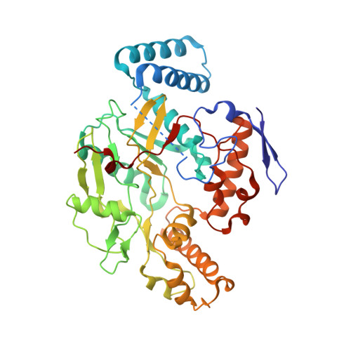
    Crystal structures are reported for the endothelial nitric oxide synthase (eNOS)-arginine-CO ternary complex as well as the neuronal nitric oxide synthase (nNOS) heme domain complexed with L: -arginine and diatomic ligands, CO or NO, in the presence of the native cofactor, tetrahydrobiopterin, or its oxidized analogs, dihydrobiopterin and 4-aminobiopterin. The nature of the biopterin has no influence on the diatomic ligand binding. The binding geometries of diatomic ligands to nitric oxide synthase (NOS) follow the {MXY}(n) formalism developed from the inorganic diatomic-metal complexes. The structures reveal some subtle structural differences between eNOS and nNOS when CO is bound to the heme which correlate well with the differences in CO stretching frequencies observed by resonance Raman techniques. The detailed hydrogen-bonding geometries depicted in the active site of nNOS structures indicate that it is the ordered active-site water molecule rather than the substrate itself that would most likely serve as a direct proton donor to the diatomic ligands (CO, NO, as well as O(2)) bound to the heme. This has important implications for the oxygen activation mechanism critical to NOS catalysis.


  • Organizational Affiliation

    Department of Molecular Biology and Biochemistry, Center in Chemical and Structural Biology, University of California, Irvine, CA 92697-3900, USA.


Macromolecules
Find similar proteins by:  (by identity cutoff)  |  3D Structure
Entity ID: 1
MoleculeChains Sequence LengthOrganismDetailsImage
Nitric-oxide synthase, brain
A, B
420Rattus norvegicusMutation(s): 0 
Gene Names: Nos1Bnos
EC: 1.14.13.39
UniProt
Find proteins for P29476 (Rattus norvegicus)
Explore P29476 
Go to UniProtKB:  P29476
Entity Groups  
Sequence Clusters30% Identity50% Identity70% Identity90% Identity95% Identity100% Identity
UniProt GroupP29476
Sequence Annotations
Expand
  • Reference Sequence
Small Molecules
Ligands 6 Unique
IDChains Name / Formula / InChI Key2D Diagram3D Interactions
HEM
Query on HEM

Download Ideal Coordinates CCD File 
E [auth A],
J [auth B]
PROTOPORPHYRIN IX CONTAINING FE
C34 H32 Fe N4 O4
KABFMIBPWCXCRK-RGGAHWMASA-L
HBI
Query on HBI

Download Ideal Coordinates CCD File 
F [auth A],
K [auth B]
7,8-DIHYDROBIOPTERIN
C9 H13 N5 O3
FEMXZDUTFRTWPE-DZSWIPIPSA-N
ARG
Query on ARG

Download Ideal Coordinates CCD File 
G [auth A],
L [auth B]
ARGININE
C6 H15 N4 O2
ODKSFYDXXFIFQN-BYPYZUCNSA-O
ZN
Query on ZN

Download Ideal Coordinates CCD File 
D [auth A]ZINC ION
Zn
PTFCDOFLOPIGGS-UHFFFAOYSA-N
ACT
Query on ACT

Download Ideal Coordinates CCD File 
C [auth A],
I [auth B]
ACETATE ION
C2 H3 O2
QTBSBXVTEAMEQO-UHFFFAOYSA-M
NO
Query on NO

Download Ideal Coordinates CCD File 
H [auth A],
M [auth B]
NITRIC OXIDE
N O
ODUCDPQEXGNKDN-UHFFFAOYSA-N
Experimental Data & Validation

Experimental Data

  • Method: X-RAY DIFFRACTION
  • Resolution: 2.05 Å
  • R-Value Free: 0.272 
  • R-Value Work: 0.225 
  • R-Value Observed: 0.225 
  • Space Group: P 21 21 21
Unit Cell:
Length ( Å )Angle ( ˚ )
a = 52.03α = 90
b = 111.16β = 90
c = 164.49γ = 90
Software Package:
Software NamePurpose
CNSrefinement
HKL-2000data reduction
SCALEPACKdata scaling
CNSphasing

Structure Validation

View Full Validation Report


Ligand Structure Quality Assessment 


Entry History 

Deposition Data

Revision History  (Full details and data files)

  • Version 1.0: 2006-08-08
    Type: Initial release
  • Version 1.1: 2008-05-01
    Changes: Version format compliance
  • Version 1.2: 2011-07-13
    Changes: Version format compliance
  • Version 1.3: 2023-08-30
    Changes: Data collection, Database references, Derived calculations, Refinement description