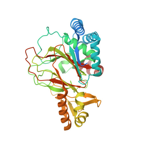
2OG7

Cystal structure of asparagine oxygenase in complex with Fe(II), 2S,3S-3-hydroxyasparagine and succinate


Experimental Data Snapshot

  • Method: X-RAY DIFFRACTION
  • Resolution: 1.66 Å
  • R-Value Free: 0.184 
  • R-Value Work: 0.168 
  • R-Value Observed: 0.168 

wwPDB Validation   3D Report Full Report


This is version 1.4 of the entry. See complete history


Literature

Mechanistic and structural basis of stereospecific Cbeta-hydroxylation in calcium-dependent antibiotic, a daptomycin-type lipopeptide

Strieker, M.Kopp, F.Mahlert, C.Essen, L.-O.Marahiel, M.A.

(2007) ACS Chem Biol 2: 187-196

  • DOI: https://doi.org/10.1021/cb700012y
  • Primary Citation of Related Structures:  
    2OG5, 2OG6, 2OG7

  • PubMed Abstract: 

    Non-ribosomally synthesized lipopeptide antibiotics of the daptomycin type are known to contain unnatural beta-modified amino acids, which are essential for bioactivity. Here we present the biochemical and structural basis for the incorporation of 3-hydroxyasparagine at position 9 in the 11-residue acidic lipopeptide lactone calcium-dependent antibiotic (CDA). Direct hydroxylation of l-asparagine by AsnO, a non-heme Fe(2+)/alpha-ketoglutarate-dependent oxygenase encoded by the CDA biosynthesis gene cluster, was validated by Fmoc derivatization of the reaction product and LC/MS analysis. The 1.45, 1.92, and 1.66 A crystal structures of AsnO as apoprotein, Fe(2+) complex, and product complex, respectively, with (2S,3S)-3-hydroxyasparagine and succinate revealed the stereoselectivity and substrate specificity of AsnO. The comparison of native and product-complex structures of AsnO showed a lid-like region (residues F208-E223) that seals the active site upon substrate binding and shields it from sterically demanding peptide substrates. Accordingly, beta-hydroxylated asparagine is synthesized prior to its incorporation into the growing CDA peptide. The AsnO structure could serve as a template for engineering novel enzymes for the synthesis of beta-hydroxylated amino acids.


  • Organizational Affiliation

    Department of Chemistry/Biochemistry, Philipps University of Marburg, Marburg, Germany.


Macromolecules
Find similar proteins by:  (by identity cutoff)  |  3D Structure
Entity ID: 1
MoleculeChains Sequence LengthOrganismDetailsImage
asparagine oxygenase357Streptomyces coelicolor A3(2)Mutation(s): 0 
Gene Names: sco3236 (asnO)
UniProt
Find proteins for Q9Z4Z5 (Streptomyces coelicolor (strain ATCC BAA-471 / A3(2) / M145))
Explore Q9Z4Z5 
Go to UniProtKB:  Q9Z4Z5
Entity Groups  
Sequence Clusters30% Identity50% Identity70% Identity90% Identity95% Identity100% Identity
UniProt GroupQ9Z4Z5
Sequence Annotations
Expand
  • Reference Sequence
Experimental Data & Validation

Experimental Data

  • Method: X-RAY DIFFRACTION
  • Resolution: 1.66 Å
  • R-Value Free: 0.184 
  • R-Value Work: 0.168 
  • R-Value Observed: 0.168 
  • Space Group: P 31 2 1
Unit Cell:
Length ( Å )Angle ( ˚ )
a = 91.308α = 90
b = 91.308β = 90
c = 90.867γ = 120
Software Package:
Software NamePurpose
REFMACrefinement
PDB_EXTRACTdata extraction
MAR345data collection
MOSFLMdata reduction
CCP4data scaling
MOLREPphasing

Structure Validation

View Full Validation Report


Entry History 

Deposition Data

Revision History  (Full details and data files)

  • Version 1.0: 2007-03-06
    Type: Initial release
  • Version 1.1: 2008-05-01
    Changes: Version format compliance
  • Version 1.2: 2011-07-13
    Changes: Advisory, Version format compliance
  • Version 1.3: 2017-10-18
    Changes: Refinement description
  • Version 1.4: 2023-08-30
    Changes: Data collection, Database references, Derived calculations, Refinement description