2Q6N

Structure of Cytochrome P450 2B4 with Bound 1-(4-cholorophenyl)imidazole


Experimental Data Snapshot

  • Method: X-RAY DIFFRACTION
  • Resolution: 3.20 Å
  • R-Value Free: 0.311 
  • R-Value Work: 0.233 

wwPDB Validation   3D Report Full Report


Ligand Structure Quality Assessment 


This is version 1.3 of the entry. See complete history


Literature

Structural and thermodynamic consequences of 1-(4-chlorophenyl)imidazole binding to cytochrome P450 2B4.

Zhao, Y.Sun, L.Muralidhara, B.K.Kumar, S.White, M.A.Stout, C.D.Halpert, J.R.

(2007) Biochemistry 46: 11559-11567

  • DOI: https://doi.org/10.1021/bi7011614
  • Primary Citation of Related Structures:  
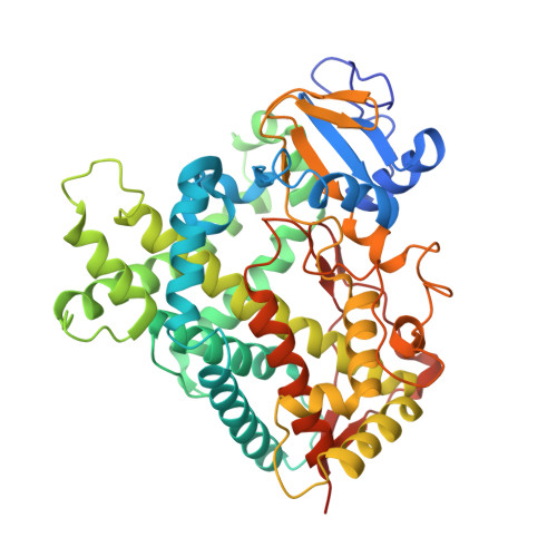
    2Q6N

  • PubMed Abstract: 

    The crystal structure of P450 2B4 bound with 1-(4-chlorophenyl)imidazole (1-CPI) has been determined to delineate the structural basis for the observed differences in binding affinity and thermodynamics relative to 4-(4-chlorophenyl)imidazole (4-CPI). Compared with the previously reported 4-CPI complex, there is a shift in the 1-CPI complex of the protein backbone in helices F and I, repositioning the side chains of Phe-206, Phe-297, and Glu-301, and leading to significant reshaping of the active site. Phe-206 and Phe-297 exchange positions, with Phe-206 becoming a ligand-contact residue, while Glu-301, rather than hydrogen bonding to the ligand, flips away from the active site and interacts with His-172. As a result the active site volume expands from 200 A3 in the 4-CPI complex to 280 A3 in the 1-CPI complex. Based on the two structures, it was predicted that a Phe-206-->Ala substitution would alter 1-CPI but not 4-CPI binding. Isothermal titration calorimetry experiments indicated that this substitution had no effect on the thermodynamic signature of 4-CPI binding to 2B4. In contrast, relative to wild-type 1-CPI binding to F206A showed significantly less favorable entropy but more favorable enthalpy. This result is consistent with loss of the aromatic side chain and possible ordering of water molecules, now able to interact with Glu-301 and exposed residues in the I-helix. Hence, thermodynamic measurements support the active site rearrangement observed in the crystal structure of the 1-CPI complex and illustrate the malleability of the active site with the fine-tuning of residue orientations and thermodynamic signatures.


  • Organizational Affiliation

    Department of Pharmacology and Toxicology, University of Texas Medical Branch, 301 University Boulevard, Galveston, Texas 77555-1031, USA.


Macromolecules
Find similar proteins by:  (by identity cutoff)  |  3D Structure
Entity ID: 1
MoleculeChains Sequence LengthOrganismDetailsImage
Cytochrome P450 2B4
A, B, C, D, E
A, B, C, D, E, F, G
478Oryctolagus cuniculusMutation(s): 10 
Gene Names: CYP2B4
EC: 1.14.14.1
Membrane Entity: Yes 
UniProt
Find proteins for P00178 (Oryctolagus cuniculus)
Explore P00178 
Go to UniProtKB:  P00178
Entity Groups  
Sequence Clusters30% Identity50% Identity70% Identity90% Identity95% Identity100% Identity
UniProt GroupP00178
Sequence Annotations
Expand
  • Reference Sequence
Small Molecules
Ligands 2 Unique
IDChains Name / Formula / InChI Key2D Diagram3D Interactions
HEM
Query on HEM

Download Ideal Coordinates CCD File 
H [auth A]
J [auth B]
L [auth C]
N [auth D]
P [auth E]
H [auth A],
J [auth B],
L [auth C],
N [auth D],
P [auth E],
R [auth F],
T [auth G]
PROTOPORPHYRIN IX CONTAINING FE
C34 H32 Fe N4 O4
KABFMIBPWCXCRK-RGGAHWMASA-L
1CI
Query on 1CI

Download Ideal Coordinates CCD File 
I [auth A]
K [auth B]
M [auth C]
O [auth D]
Q [auth E]
I [auth A],
K [auth B],
M [auth C],
O [auth D],
Q [auth E],
S [auth F],
U [auth G]
1-(4-CHLOROPHENYL)-1H-IMIDAZOLE
C9 H7 Cl N2
BARLRKAYTDVUIS-UHFFFAOYSA-N
Binding Affinity Annotations 
IDSourceBinding Affinity
1CI BindingDB:  2Q6N -TΔS: min: 0.42, max: 13.8 (kJ/mol) from 2 assay(s)
Experimental Data & Validation

Experimental Data

  • Method: X-RAY DIFFRACTION
  • Resolution: 3.20 Å
  • R-Value Free: 0.311 
  • R-Value Work: 0.233 
  • Space Group: P 21 21 21
Unit Cell:
Length ( Å )Angle ( ˚ )
a = 140α = 90
b = 147.31β = 90
c = 238.49γ = 90
Software Package:
Software NamePurpose
SCALAdata scaling
PHASERphasing
CNSrefinement
PDB_EXTRACTdata extraction
MOSFLMdata reduction

Structure Validation

View Full Validation Report


Ligand Structure Quality Assessment 


Entry History 

Deposition Data

Revision History  (Full details and data files)

  • Version 1.0: 2007-11-06
    Type: Initial release
  • Version 1.1: 2011-07-13
    Changes: Version format compliance
  • Version 1.2: 2021-10-20
    Changes: Database references, Derived calculations
  • Version 1.3: 2023-08-30
    Changes: Data collection, Refinement description