2W3X

Crystal structure of a bifunctional hotdog fold thioesterase in enediyne biosynthesis, CalE7


Experimental Data Snapshot

  • Method: X-RAY DIFFRACTION
  • Resolution: 1.75 Å
  • R-Value Free: 0.235 
  • R-Value Work: 0.196 
  • R-Value Observed: 0.196 

wwPDB Validation   3D Report Full Report


Ligand Structure Quality Assessment 


This is version 1.3 of the entry. See complete history


Literature

Structure and Catalytic Mechanism of the Thioesterase Cale7 in Enediyne Biosynthesis.

Kotaka, M.Kong, R.Qureshi, I.Ho, Q.S.Sun, H.Liew, C.W.Goh, L.P.Cheung, P.Mu, Y.Lescar, J.Liang, Z.X.

(2009) J Biol Chem 284: 15739

  • DOI: https://doi.org/10.1074/jbc.M809669200
  • Primary Citation of Related Structures:  
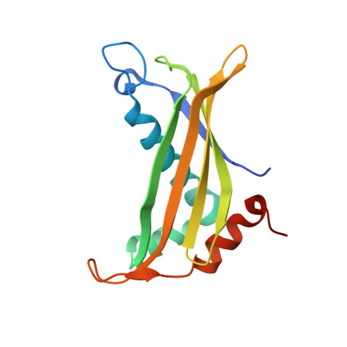
    2W3X

  • PubMed Abstract: 

    The biosynthesis of the enediyne moiety of the antitumor natural product calicheamicin involves an iterative polyketide synthase (CalE8) and other ancillary enzymes. In the proposed mechanism for the early stage of 10-membered enediyne biosynthesis, CalE8 produces a carbonyl-conjugated polyene with the assistance of a putative thioesterase (CalE7). We have determined the x-ray crystal structure of CalE7 and found that the subunit adopts a hotdog fold with an elongated and kinked substrate-binding channel embedded between two subunits. The 1.75-A crystal structure revealed that CalE7 does not contain a critical catalytic residue (Glu or Asp) conserved in other hotdog fold thioesterases. Based on biochemical and site-directed mutagenesis studies, we proposed a catalytic mechanism in which the conserved Arg(37) plays a crucial role in the hydrolysis of the thioester bond, and that Tyr(29) and a hydrogen-bonded water network assist the decarboxylation of the beta-ketocarboxylic acid intermediate. Moreover, computational docking suggested that the substrate-binding channel binds a polyene substrate that contains a single cis double bond at the C4/C5 position, raising the possibility that the C4=C5 double bond in the enediyne moiety could be generated by the iterative polyketide synthase. Together, the results revealed a hotdog fold thioesterase distinct from the common type I and type II thioesterases associated with polyketide biosynthesis and provided interesting insight into the enediyne biosynthetic mechanism.


  • Organizational Affiliation

    School of Biological Sciences, Nanyang Technological University, 60 Nanyang Drive, Singapore 637551, Singapore.


Macromolecules
Find similar proteins by:  (by identity cutoff)  |  3D Structure
Entity ID: 1
MoleculeChains Sequence LengthOrganismDetailsImage
CALE7
A, B, C, D, E
A, B, C, D, E, F
147Micromonospora echinosporaMutation(s): 0 
UniProt
Find proteins for Q8KNG2 (Micromonospora echinospora)
Explore Q8KNG2 
Go to UniProtKB:  Q8KNG2
Entity Groups  
Sequence Clusters30% Identity50% Identity70% Identity90% Identity95% Identity100% Identity
UniProt GroupQ8KNG2
Sequence Annotations
Expand
  • Reference Sequence
Small Molecules
Ligands 4 Unique
IDChains Name / Formula / InChI Key2D Diagram3D Interactions
JEF
Query on JEF

Download Ideal Coordinates CCD File 
H [auth A]O-(O-(2-AMINOPROPYL)-O'-(2-METHOXYETHYL)POLYPROPYLENE GLYCOL 500)
C30 H63 N O10
ICCXIDTYQFYPNV-RUMGZKRTSA-N
POP
Query on POP

Download Ideal Coordinates CCD File 
J [auth C],
M [auth D]
PYROPHOSPHATE 2-
H2 O7 P2
XPPKVPWEQAFLFU-UHFFFAOYSA-L
PO4
Query on PO4

Download Ideal Coordinates CCD File 
G [auth A],
I [auth B]
PHOSPHATE ION
O4 P
NBIIXXVUZAFLBC-UHFFFAOYSA-K
GOL
Query on GOL

Download Ideal Coordinates CCD File 
K [auth C],
L [auth C]
GLYCEROL
C3 H8 O3
PEDCQBHIVMGVHV-UHFFFAOYSA-N
Experimental Data & Validation

Experimental Data

  • Method: X-RAY DIFFRACTION
  • Resolution: 1.75 Å
  • R-Value Free: 0.235 
  • R-Value Work: 0.196 
  • R-Value Observed: 0.196 
  • Space Group: C 1 2 1
Unit Cell:
Length ( Å )Angle ( ˚ )
a = 101.95α = 90
b = 73.32β = 98.69
c = 124.41γ = 90
Software Package:
Software NamePurpose
CNSrefinement
DENZOdata reduction
SCALEPACKdata scaling
MrBUMPphasing

Structure Validation

View Full Validation Report


Ligand Structure Quality Assessment 


Entry History 

Deposition Data

Revision History  (Full details and data files)

  • Version 1.0: 2009-04-07
    Type: Initial release
  • Version 1.1: 2011-05-08
    Changes: Version format compliance
  • Version 1.2: 2011-07-13
    Changes: Version format compliance
  • Version 1.3: 2023-12-13
    Changes: Data collection, Database references, Derived calculations, Other, Refinement description