2WOD

Crystal Structure of the dinitrogenase reductase-activating glycohydrolase (DRAG) from Rhodospirillum rubrum in complex with ADP- ribsoyllysine


Experimental Data Snapshot

  • Method: X-RAY DIFFRACTION
  • Resolution: 2.25 Å
  • R-Value Free: 0.242 
  • R-Value Work: 0.197 
  • R-Value Observed: 0.199 

wwPDB Validation   3D Report Full Report


Ligand Structure Quality Assessment 


This is version 1.2 of the entry. See complete history


Literature

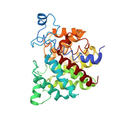
Mechanism of Adp-Ribosylation Removal Revealed by the Structure and Ligand Complexes of the Dimanganese Mono-Adp-Ribosylhydrolase Drag.

Berthold, C.L.Wang, H.Nordlund, S.Hogbom, M.

(2009) Proc Natl Acad Sci U S A 106: 14247

  • DOI: https://doi.org/10.1073/pnas.0905906106
  • Primary Citation of Related Structures:  
    2WOC, 2WOD, 2WOE

  • PubMed Abstract: 

    ADP-ribosylation is a ubiquitous regulatory posttranslational modification involved in numerous key processes such as DNA repair, transcription, cell differentiation, apoptosis, and the pathogenic mechanism of certain bacterial toxins. Despite the importance of this reversible process, very little is known about the structure and mechanism of the hydrolases that catalyze removal of the ADP-ribose moiety. In the phototrophic bacterium Rhodospirillum rubrum, dinitrogenase reductase-activating glycohydrolase (DraG), a dimanganese enzyme that reversibly associates with the cell membrane, is a key player in the regulation of nitrogenase activity. DraG has long served as a model protein for ADP-ribosylhydrolases. Here, we present the crystal structure of DraG in the holo and ADP-ribose bound forms. We also present the structure of a reaction intermediate analogue and propose a detailed catalytic mechanism for protein de-ADP-ribosylation involving ring opening of the substrate ribose. In addition, the particular manganese coordination in DraG suggests a rationale for the enzyme's preference for manganese over magnesium, although not requiring a redox active metal for the reaction.


  • Organizational Affiliation

    Department of Biochemistry and Biophysics, Stockholm Center for Biomembrane Research, Arrhenius Laboratories for Natural Sciences, Stockholm University, SE-10691 Stockholm, Sweden.


Macromolecules
Find similar proteins by:  (by identity cutoff)  |  3D Structure
Entity ID: 1
MoleculeChains Sequence LengthOrganismDetailsImage
ADP-RIBOSYL-[DINITROGEN REDUCTASE] GLYCOHYDROLASE
A, B
299Rhodospirillum rubrumMutation(s): 0 
EC: 3.2.2.24
UniProt
Find proteins for P14300 (Rhodospirillum rubrum)
Explore P14300 
Go to UniProtKB:  P14300
Entity Groups  
Sequence Clusters30% Identity50% Identity70% Identity90% Identity95% Identity100% Identity
UniProt GroupP14300
Sequence Annotations
Expand
  • Reference Sequence
Small Molecules
Ligands 4 Unique
IDChains Name / Formula / InChI Key2D Diagram3D Interactions
ZZC
Query on ZZC

Download Ideal Coordinates CCD File 
D [auth A],
F [auth B]
[(2R,3S,4R,5R)-5-(6-AMINOPURIN-9-YL)-3,4-DIHYDROXY-OXOLAN-2-YL]METHYL [[(2R,3R)-2,3-DIHYDROXY-4-OXO-PENTOXY]-OXIDO-PHOSPHORYL] PHOSPHATE
C15 H21 N5 O13 P2
IZTPERUWNZCNNH-GHVQHMAVSA-L
GOL
Query on GOL

Download Ideal Coordinates CCD File 
H [auth B],
I [auth B]
GLYCEROL
C3 H8 O3
PEDCQBHIVMGVHV-UHFFFAOYSA-N
MN
Query on MN

Download Ideal Coordinates CCD File 
C [auth A],
E [auth B]
MANGANESE (II) ION
Mn
WAEMQWOKJMHJLA-UHFFFAOYSA-N
CL
Query on CL

Download Ideal Coordinates CCD File 
G [auth B]CHLORIDE ION
Cl
VEXZGXHMUGYJMC-UHFFFAOYSA-M
Experimental Data & Validation

Experimental Data

  • Method: X-RAY DIFFRACTION
  • Resolution: 2.25 Å
  • R-Value Free: 0.242 
  • R-Value Work: 0.197 
  • R-Value Observed: 0.199 
  • Space Group: P 31 2 1
Unit Cell:
Length ( Å )Angle ( ˚ )
a = 77.306α = 90
b = 77.306β = 90
c = 245.593γ = 120
Software Package:
Software NamePurpose
REFMACrefinement
MOSFLMdata reduction
SCALAdata scaling
MOLREPphasing

Structure Validation

View Full Validation Report


Ligand Structure Quality Assessment 


Entry History 

Deposition Data

Revision History  (Full details and data files)

  • Version 1.0: 2009-08-11
    Type: Initial release
  • Version 1.1: 2011-07-13
    Changes: Advisory, Version format compliance
  • Version 1.2: 2018-01-17
    Changes: Data collection