2Z2P

Crystal Structure of catalytically inactive H270A virginiamycin B lyase from Staphylococcus aureus with Quinupristin


Experimental Data Snapshot

  • Method: X-RAY DIFFRACTION
  • Resolution: 2.80 Å
  • R-Value Free: 0.318 
  • R-Value Work: 0.262 
  • R-Value Observed: 0.268 

wwPDB Validation   3D Report Full Report


Ligand Structure Quality Assessment 


This is version 2.2 of the entry. See complete history


Literature

Structural Basis for Streptogramin B Resistance in Staphylococcus Aureus by Virginiamycin B Lyase

Korczynska, M.Mukhtar, T.A.Wright, G.D.Berghuis, A.M.

(2007) Proc Natl Acad Sci U S A 104: 10388

  • DOI: https://doi.org/10.1073/pnas.0701809104
  • Primary Citation of Related Structures:  
    2Z2N, 2Z2O, 2Z2P

  • PubMed Abstract: 

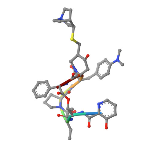
    The streptogramin combination therapy of quinupristin-dalfopristin (Synercid) is used to treat infections caused by bacterial pathogens, such as methicillin-resistant Staphylococcus aureus and vancomycin-resistant Enterococcus faecium. However, the effectiveness of this therapy is being compromised because of an increased incidence of streptogramin resistance. One of the clinically observed mechanisms of resistance is enzymatic inactivation of the type B streptogramins, such as quinupristin, by a streptogramin B lyase, i.e., virginiamycin B lyase (Vgb). The enzyme catalyzes the linearization of the cyclic antibiotic via a cleavage that requires a divalent metal ion. Here, we present crystal structures of Vgb from S. aureus in its apoenzyme form and in complex with quinupristin and Mg2+ at 1.65- and 2.8-A resolution, respectively. The fold of the enzyme is that of a seven-bladed beta-propeller, although the sequence reveals no similarity to other known members of this structural family. Quinupristin binds to a large depression on the surface of the enzyme, where it predominantly forms van der Waals interactions. Validated by site-directed mutagenesis studies, a reaction mechanism is proposed in which the initial abstraction of a proton is facilitated by a Mg2+ -linked conjugated system. Analysis of the Vgb-quinupristin structure and comparison with the complex between quinupristin and its natural target, the 50S ribosomal subunit, reveals features that can be exploited for developing streptogramins that are impervious to Vgb-mediated resistance.


  • Organizational Affiliation

    Department of Biochemistry, McGill University, Montreal, QC, Canada H3A 4A2.


Macromolecules
Find similar proteins by:  (by identity cutoff)  |  3D Structure
Entity ID: 1
MoleculeChains Sequence LengthOrganismDetailsImage
Virginiamycin B lyase
A, B
299Staphylococcus aureusMutation(s): 1 
Gene Names: vgbvgh
EC: 4.2.99
UniProt
Find proteins for P17978 (Staphylococcus aureus)
Explore P17978 
Go to UniProtKB:  P17978
Entity Groups  
Sequence Clusters30% Identity50% Identity70% Identity90% Identity95% Identity100% Identity
UniProt GroupP17978
Sequence Annotations
Expand
  • Reference Sequence

Find similar proteins by:  Sequence   |   3D Structure  

Entity ID: 2
MoleculeChains Sequence LengthOrganismDetailsImage
Quinupristin
C, D
8Streptomyces graminofaciensMutation(s): 0 
Sequence Annotations
Expand
  • Reference Sequence
Small Molecules
Modified Residues  1 Unique
IDChains TypeFormula2D DiagramParent
MHU
Query on MHU
C, D
L-PEPTIDE LINKINGC12 H18 N2 O2PHE
Biologically Interesting Molecules (External Reference) 1 Unique
Experimental Data & Validation

Experimental Data

  • Method: X-RAY DIFFRACTION
  • Resolution: 2.80 Å
  • R-Value Free: 0.318 
  • R-Value Work: 0.262 
  • R-Value Observed: 0.268 
  • Space Group: P 21 21 21
Unit Cell:
Length ( Å )Angle ( ˚ )
a = 70.999α = 90
b = 93.7β = 90
c = 95.301γ = 90
Software Package:
Software NamePurpose
PHASERphasing
REFMACrefinement
HKL-2000data reduction
HKL-2000data scaling

Structure Validation

View Full Validation Report


Ligand Structure Quality Assessment 


Entry History 

Deposition Data

Revision History  (Full details and data files)

  • Version 1.0: 2007-06-19
    Type: Initial release
  • Version 1.1: 2011-06-14
    Changes: Version format compliance
  • Version 1.2: 2011-07-13
    Changes: Version format compliance
  • Version 1.3: 2011-07-27
    Changes: Advisory, Atomic model, Database references, Derived calculations, Non-polymer description, Structure summary
  • Version 1.4: 2012-12-12
    Changes: Other
  • Version 1.5: 2015-04-22
    Changes: Database references
  • Version 1.6: 2017-11-01
    Changes: Derived calculations
  • Version 2.0: 2023-07-12
    Changes: Advisory, Atomic model, Data collection, Database references, Derived calculations, Polymer sequence, Refinement description, Source and taxonomy, Structure summary
  • Version 2.1: 2023-11-08
    Changes: Data collection, Refinement description
  • Version 2.2: 2023-11-15
    Changes: Data collection, Derived calculations