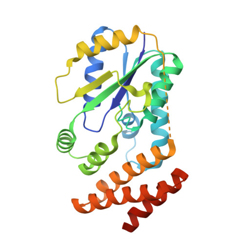
3A4M

Crystal structure of archaeal O-phosphoseryl-tRNA(Sec) kinase


Experimental Data Snapshot

  • Method: X-RAY DIFFRACTION
  • Resolution: 1.79 Å
  • R-Value Free: 0.253 
  • R-Value Work: 0.209 
  • R-Value Observed: 0.211 

wwPDB Validation   3D Report Full Report


Ligand Structure Quality Assessment 


This is version 1.2 of the entry. See complete history


Literature

Structure of a tRNA-dependent kinase essential for selenocysteine decoding

Araiso, Y.Sherrer, R.L.Ishitani, R.Ho, J.M.L.Soll, D.Nureki, O.

(2009) Proc Natl Acad Sci U S A 106: 16215-16220

  • DOI: https://doi.org/10.1073/pnas.0908861106
  • Primary Citation of Related Structures:  
    3A4L, 3A4M, 3A4N

  • PubMed Abstract: 

    Compared to bacteria, archaea and eukaryotes employ an additional enzyme for the biosynthesis of selenocysteine (Sec), the 21(st) natural amino acid (aa). An essential RNA-dependent kinase, O-phosphoseryl-tRNA(Sec) kinase (PSTK), converts seryl-tRNA(Sec) to O-phosphoseryl-tRNA(Sec), the immediate precursor of selenocysteinyl-tRNA(Sec). The sequence of Methanocaldococcus jannaschii PSTK (MjPSTK) suggests an N-terminal kinase domain (177 aa) followed by a presumed tRNA binding region (75 aa). The structures of MjPSTK complexed with ADP and AMPPNP revealed that this enzyme belongs to the P-loop kinase class, and that the kinase domain is closely related to gluconate kinase and adenylate kinase. ATP is bound by the P-loop domain (residues 11-18). Formed by antiparallel dimerization of two PSTK monomers, the enzyme structure shows a deep groove with positive electrostatic potential. Located in this groove is the enzyme's active site, which biochemical and genetic data suggest is composed of Asp-41, Arg-44, Glu-55, Tyr-82, Tyr-83, Met-86, and Met-132. Based on structural comparison with Escherichia coli adenylate kinase a docking model was generated that assigns these amino acids to the recognition of the terminal A76-Ser moieties of Ser-tRNA(Sec). The geometry and electrostatic environment of the groove in MjPSTK are perfectly complementary to the unusually long acceptor helix of tRNA(Sec).


  • Organizational Affiliation

    Department of Basic Medical Sciences, Institute of Medical Science, The University of Tokyo, 4-6-1 Shirokanedai, Minato-ku, Tokyo 108-8639, Japan.


Macromolecules
Find similar proteins by:  (by identity cutoff)  |  3D Structure
Entity ID: 1
MoleculeChains Sequence LengthOrganismDetailsImage
L-seryl-tRNA(Sec) kinase
A, B
260Methanocaldococcus jannaschiiMutation(s): 0 
Gene Names: MJ1538
EC: 2.7.1
UniProt
Find proteins for Q58933 (Methanocaldococcus jannaschii (strain ATCC 43067 / DSM 2661 / JAL-1 / JCM 10045 / NBRC 100440))
Explore Q58933 
Go to UniProtKB:  Q58933
Entity Groups  
Sequence Clusters30% Identity50% Identity70% Identity90% Identity95% Identity100% Identity
UniProt GroupQ58933
Sequence Annotations
Expand
  • Reference Sequence
Small Molecules
Ligands 4 Unique
IDChains Name / Formula / InChI Key2D Diagram3D Interactions
ADP
Query on ADP

Download Ideal Coordinates CCD File 
C [auth A],
G [auth B]
ADENOSINE-5'-DIPHOSPHATE
C10 H15 N5 O10 P2
XTWYTFMLZFPYCI-KQYNXXCUSA-N
IOD
Query on IOD

Download Ideal Coordinates CCD File 
E [auth A],
I [auth B]
IODIDE ION
I
XMBWDFGMSWQBCA-UHFFFAOYSA-M
EDO
Query on EDO

Download Ideal Coordinates CCD File 
F [auth A],
J [auth B],
K [auth B],
L [auth B]
1,2-ETHANEDIOL
C2 H6 O2
LYCAIKOWRPUZTN-UHFFFAOYSA-N
MG
Query on MG

Download Ideal Coordinates CCD File 
D [auth A],
H [auth B]
MAGNESIUM ION
Mg
JLVVSXFLKOJNIY-UHFFFAOYSA-N
Experimental Data & Validation

Experimental Data

  • Method: X-RAY DIFFRACTION
  • Resolution: 1.79 Å
  • R-Value Free: 0.253 
  • R-Value Work: 0.209 
  • R-Value Observed: 0.211 
  • Space Group: P 1 21 1
Unit Cell:
Length ( Å )Angle ( ˚ )
a = 58.156α = 90
b = 74.524β = 113.37
c = 64.817γ = 90
Software Package:
Software NamePurpose
HKL-2000data collection
SHARPphasing
PHENIXrefinement
HKL-2000data reduction
HKL-2000data scaling

Structure Validation

View Full Validation Report


Ligand Structure Quality Assessment 


Entry History 

Deposition Data

Revision History  (Full details and data files)

  • Version 1.0: 2009-10-20
    Type: Initial release
  • Version 1.1: 2011-07-13
    Changes: Version format compliance
  • Version 1.2: 2024-03-13
    Changes: Data collection, Database references, Derived calculations