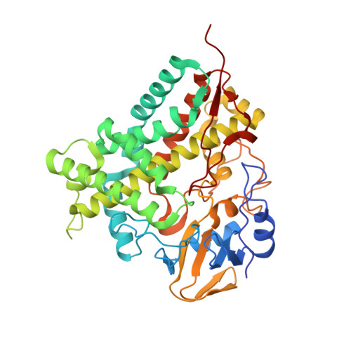
Regio- and stereospecificity of filipin hydroxylation sites revealed by crystal structures of cytochrome P450 105P1 and 105D6 from Streptomyces avermitilis
Xu, L.H., Fushinobu, S., Takamatsu, S., Wakagi, T., Ikeda, H., Shoun, H.(2010) J Biol Chem 285: 16844-16853
- PubMed: 20375018
- DOI: https://doi.org/10.1074/jbc.M109.092460
- Primary Citation of Related Structures:
3ABA, 3ABB - PubMed Abstract:
The polyene macrolide antibiotic filipin is widely used as a probe for cholesterol and a diagnostic tool for type C Niemann-Pick disease. Two position-specific P450 enzymes are involved in the post-polyketide modification of filipin during its biosynthesis, thereby providing molecular diversity to the "filipin complex." CYP105P1 and CYP105D6 from Streptomyces avermitilis, despite their high sequence similarities, catalyze filipin hydroxylation at different positions, C26 and C1', respectively. Here, we determined the crystal structure of the CYP105P1-filipin I complex. The distal pocket of CYP105P1 has the second largest size among P450 hydroxylases that act on macrolide substrates. Compared with previously determined substrate-free structures, the FG helices showed significant closing motion on substrate binding. The long BC loop region adopts a unique extended conformation without a B' helix. The binding site is essentially hydrophobic, but numerous water molecules are involved in recognizing the polyol side of the substrate. Therefore, the distal pocket of CYP105P1 provides a specific environment for the large filipin substrate to bind with its pro-S side of position C26 directed toward the heme iron. The ligand-free CYP105D6 structure was also determined. A small sub-pocket accommodating the long alkyl side chain of filipin I was observed in the CYP105P1 structure but was absent in the CYP105D6 structure, indicating that filipin cannot bind to CYP105D6 with a similar orientation due to steric hindrance. This observation can explain the strict regiospecificity of these enzymes.
Organizational Affiliation:
Department of Biotechnology, Graduate School of Agriculture and Life Sciences, the University of Tokyo, 1-1-1 Yayoi, Bunkyo-ku, Tokyo 113-8657, Japan.