3DCO

Drosophila NOD (3DC4) and Bovine Tubulin (1JFF) Docked into the 11-Angstrom Cryo-EM Map of Nucleotide-Free NOD Complexed to the Microtubule


Experimental Data Snapshot

  • Method: ELECTRON MICROSCOPY
  • Resolution: 11.0 Å
  • Aggregation State: HELICAL ARRAY 
  • Reconstruction Method: SINGLE PARTICLE 

wwPDB Validation   3D Report Full Report


This is version 1.4 of the entry. See complete history


Literature

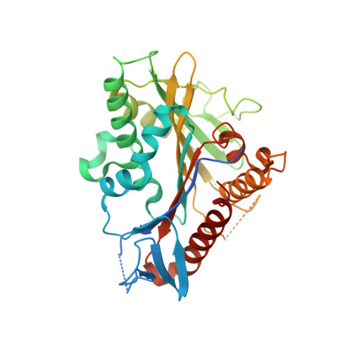
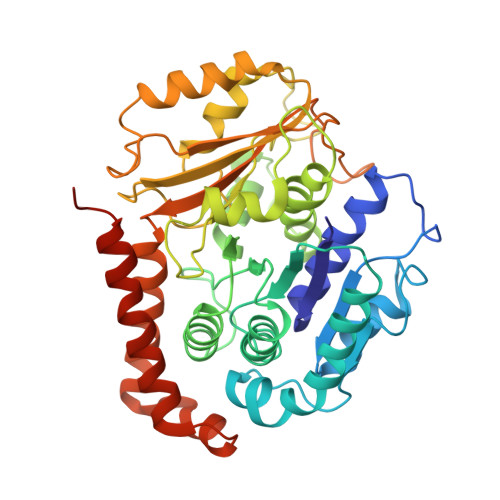
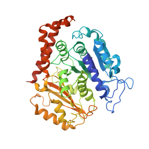
ATPase cycle of the nonmotile kinesin NOD allows microtubule end tracking and drives chromosome movement.

Cochran, J.C.Sindelar, C.V.Mulko, N.K.Collins, K.A.Kong, S.E.Hawley, R.S.Kull, F.J.

(2009) Cell 136: 110-122

  • DOI: https://doi.org/10.1016/j.cell.2008.11.048
  • Primary Citation of Related Structures:  
    3DC4, 3DCB, 3DCO

  • PubMed Abstract: 

    Segregation of nonexchange chromosomes during Drosophila melanogaster meiosis requires the proper function of NOD, a nonmotile kinesin-10. We have determined the X-ray crystal structure of the NOD catalytic domain in the ADP- and AMPPNP-bound states. These structures reveal an alternate conformation of the microtubule binding region as well as a nucleotide-sensitive relay of hydrogen bonds at the active site. Additionally, a cryo-electron microscopy reconstruction of the nucleotide-free microtubule-NOD complex shows an atypical binding orientation. Thermodynamic studies show that NOD binds tightly to microtubules in the nucleotide-free state, yet other nucleotide states, including AMPPNP, are weakened. Our pre-steady-state kinetic analysis demonstrates that NOD interaction with microtubules occurs slowly with weak activation of ADP product release. Upon rapid substrate binding, NOD detaches from the microtubule prior to the rate-limiting step of ATP hydrolysis, which is also atypical for a kinesin. We propose a model for NOD's microtubule plus-end tracking that drives chromosome movement.


  • Organizational Affiliation

    Department of Chemistry, Dartmouth College, Hanover, NH 03755, USA.


Macromolecules
Find similar proteins by:  (by identity cutoff)  |  3D Structure
Entity ID: 1
MoleculeChains Sequence LengthOrganismDetailsImage
Kinesin-like protein NodA [auth N]344Drosophila melanogasterMutation(s): 0 
Gene Names: CG1763nodNODA
UniProt
Find proteins for P18105 (Drosophila melanogaster)
Explore P18105 
Go to UniProtKB:  P18105
Entity Groups  
Sequence Clusters30% Identity50% Identity70% Identity90% Identity95% Identity100% Identity
UniProt GroupP18105
Sequence Annotations
Expand
  • Reference Sequence
Find similar proteins by:  (by identity cutoff)  |  3D Structure
Entity ID: 2
MoleculeChains Sequence LengthOrganismDetailsImage
Bovine Alpha TubulinB [auth A]451Bos taurusMutation(s): 0 
UniProt
Find proteins for P81947 (Bos taurus)
Explore P81947 
Go to UniProtKB:  P81947
Entity Groups  
Sequence Clusters30% Identity50% Identity70% Identity90% Identity95% Identity100% Identity
UniProt GroupP81947
Sequence Annotations
Expand
  • Reference Sequence
Find similar proteins by:  (by identity cutoff)  |  3D Structure
Entity ID: 3
MoleculeChains Sequence LengthOrganismDetailsImage
Bovine Beta TubulinC [auth B]445Bos taurusMutation(s): 0 
UniProt
Find proteins for Q6B856 (Bos taurus)
Explore Q6B856 
Go to UniProtKB:  Q6B856
Entity Groups  
Sequence Clusters30% Identity50% Identity70% Identity90% Identity95% Identity100% Identity
UniProt GroupQ6B856
Sequence Annotations
Expand
  • Reference Sequence
Small Molecules
Ligands 6 Unique
IDChains Name / Formula / InChI Key2D Diagram3D Interactions
TA1
Query on TA1

Download Ideal Coordinates CCD File 
J [auth B]TAXOL
C47 H51 N O14
RCINICONZNJXQF-MZXODVADSA-N
GTP
Query on GTP

Download Ideal Coordinates CCD File 
H [auth B]GUANOSINE-5'-TRIPHOSPHATE
C10 H16 N5 O14 P3
XKMLYUALXHKNFT-UUOKFMHZSA-N
GDP
Query on GDP

Download Ideal Coordinates CCD File 
I [auth B]GUANOSINE-5'-DIPHOSPHATE
C10 H15 N5 O11 P2
QGWNDRXFNXRZMB-UUOKFMHZSA-N
ADP
Query on ADP

Download Ideal Coordinates CCD File 
E [auth N]ADENOSINE-5'-DIPHOSPHATE
C10 H15 N5 O10 P2
XTWYTFMLZFPYCI-KQYNXXCUSA-N
ZN
Query on ZN

Download Ideal Coordinates CCD File 
G [auth A]ZINC ION
Zn
PTFCDOFLOPIGGS-UHFFFAOYSA-N
MG
Query on MG

Download Ideal Coordinates CCD File 
D [auth N],
F [auth A]
MAGNESIUM ION
Mg
JLVVSXFLKOJNIY-UHFFFAOYSA-N
Binding Affinity Annotations 
IDSourceBinding Affinity
TA1 BindingDB:  3DCO EC50: 520 (nM) from 1 assay(s)
Experimental Data & Validation

Experimental Data

  • Method: ELECTRON MICROSCOPY
  • Resolution: 11.0 Å
  • Aggregation State: HELICAL ARRAY 
  • Reconstruction Method: SINGLE PARTICLE 
EM Software:
TaskSoftware PackageVersion
RECONSTRUCTIONSPIDER

Structure Validation

View Full Validation Report


Entry History 

Deposition Data

Revision History  (Full details and data files)

  • Version 1.0: 2009-02-10
    Type: Initial release
  • Version 1.1: 2011-07-13
    Changes: Version format compliance
  • Version 1.2: 2016-11-09
    Changes: Other
  • Version 1.3: 2018-07-18
    Changes: Author supporting evidence, Data collection
  • Version 1.4: 2024-02-21
    Changes: Data collection, Database references, Derived calculations, Refinement description