3DY0

Crystal Structure of Cleaved PCI Bound to Heparin


Experimental Data Snapshot

  • Method: X-RAY DIFFRACTION
  • Resolution: 1.55 Å
  • R-Value Free: 0.217 
  • R-Value Work: 0.197 
  • R-Value Observed: 0.197 

wwPDB Validation   3D Report Full Report


This is version 2.1 of the entry. See complete history


Literature

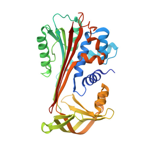
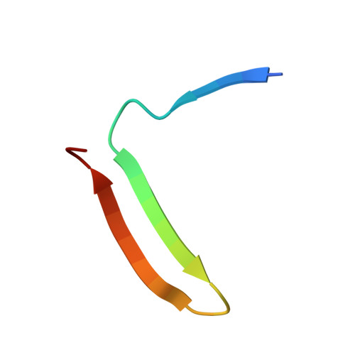
The heparin binding site of protein C inhibitor is protease-dependent.

Li, W.Huntington, J.A.

(2008) J Biol Chem 283: 36039-36045

  • DOI: https://doi.org/10.1074/jbc.M805974200
  • Primary Citation of Related Structures:  
    3DY0

  • PubMed Abstract: 

    Protein C inhibitor (PCI) is a member of the serpin family of protease inhibitors with many biological functions and broad inhibitory specificity. Its major targets in blood are thrombin and activated protein C (APC), and the inhibition of both enzymes can be accelerated by glycosaminoglycans, including heparin. Acceleration of thrombin and APC inhibition by PCI requires that both protease and inhibitor bind to the same heparin chain to form a bridged Michaelis complex. However, the position of the heparin binding site of APC is opposite to that of thrombin, and formation of the bridged complexes must require either radical reorientation of the proteases relative to PCI or alternate heparin binding modes for PCI. In this study, we investigate how heparin bridges thrombin and APC to PCI by determining the effect of mutations in and around the putative heparin binding site of PCI. We found that heparin binds PCI in a linear fashion along helix H to bridge thrombin, consistent with our recent crystal structure (3B9F), but that it must rotate by approximately 60 degrees to engage Arg-229 to bridge APC. To gain insight into the possible modes of heparin binding to PCI, we solved a crystal structure of cleaved PCI bound to an octasaccharide heparin fragment to 1.55 angstroms resolution. The structure reveals a binding mode across the N terminus of helix H to engage Arg-229 and align the heparin binding site of APC. A molecular model for the heparin-bridged PCI.APC complex was built based on mutagenesis and structural data.


  • Organizational Affiliation

    Department of Haematology, University of Cambridge, Cambridge Institute for Medical Research, Wellcome Trust/MRC Building, Hills Road, Cambridge, CB2 0XY, United Kingdom.


Macromolecules
Find similar proteins by:  (by identity cutoff)  |  3D Structure
Entity ID: 1
MoleculeChains Sequence LengthOrganismDetailsImage
N-terminus Plasma serine protease inhibitor336Homo sapiensMutation(s): 0 
Gene Names: SERPINA5PCIPLANH3PROCI
UniProt & NIH Common Fund Data Resources
Find proteins for P05154 (Homo sapiens)
Explore P05154 
Go to UniProtKB:  P05154
PHAROS:  P05154
GTEx:  ENSG00000188488 
Entity Groups  
Sequence Clusters30% Identity50% Identity70% Identity90% Identity95% Identity100% Identity
UniProt GroupP05154
Sequence Annotations
Expand
  • Reference Sequence
Find similar proteins by:  (by identity cutoff)  |  3D Structure
Entity ID: 2
MoleculeChains Sequence LengthOrganismDetailsImage
C-terminus Plasma serine protease inhibitor29Homo sapiensMutation(s): 0 
Gene Names: SERPINA5PCIPLANH3PROCI
UniProt & NIH Common Fund Data Resources
Find proteins for P05154 (Homo sapiens)
Explore P05154 
Go to UniProtKB:  P05154
PHAROS:  P05154
GTEx:  ENSG00000188488 
Entity Groups  
Sequence Clusters30% Identity50% Identity70% Identity90% Identity95% Identity100% Identity
UniProt GroupP05154
Sequence Annotations
Expand
  • Reference Sequence
Oligosaccharides

Help

Entity ID: 3
MoleculeChains Length2D Diagram Glycosylation3D Interactions
2-O-sulfo-alpha-L-idopyranuronic acid-(1-4)-2-deoxy-6-O-sulfo-2-(sulfoamino)-alpha-D-glucopyranose-(1-4)-2-O-sulfo-alpha-L-idopyranuronic acid-(1-4)-2-deoxy-6-O-sulfo-2-(sulfoamino)-alpha-D-glucopyranose-(1-4)-2-O-sulfo-alpha-L-idopyranuronic acid
C
5N/A
Glycosylation Resources
GlyTouCan:  G93330CR
GlyCosmos:  G93330CR
Experimental Data & Validation

Experimental Data

  • Method: X-RAY DIFFRACTION
  • Resolution: 1.55 Å
  • R-Value Free: 0.217 
  • R-Value Work: 0.197 
  • R-Value Observed: 0.197 
  • Space Group: P 1 21 1
Unit Cell:
Length ( Å )Angle ( ˚ )
a = 61.051α = 90
b = 49.451β = 113.08
c = 64.969γ = 90
Software Package:
Software NamePurpose
CNSrefinement
ADSCdata collection
PHASERphasing
MOSFLMdata reduction
SCALAdata scaling

Structure Validation

View Full Validation Report


Entry History 

Deposition Data

Revision History  (Full details and data files)

  • Version 1.0: 2008-10-28
    Type: Initial release
  • Version 1.1: 2011-07-13
    Changes: Non-polymer description, Version format compliance
  • Version 2.0: 2020-07-29
    Type: Remediation
    Reason: Carbohydrate remediation
    Changes: Advisory, Atomic model, Data collection, Database references, Derived calculations, Refinement description, Structure summary
  • Version 2.1: 2023-08-30
    Changes: Data collection, Database references, Refinement description, Structure summary