3ELO

Crystal Structure of Human Pancreatic Prophospholipase A2


Experimental Data Snapshot

  • Method: X-RAY DIFFRACTION
  • Resolution: 1.55 Å
  • R-Value Free: 0.183 
  • R-Value Work: 0.145 
  • R-Value Observed: 0.147 

wwPDB Validation   3D Report Full Report


This is version 1.2 of the entry. See complete history


Literature

Structural insight into the activation mechanism of human pancreatic prophospholipase A2

Xu, W.Yi, L.Feng, Y.Chen, L.Liu, J.

(2009) J Biol Chem 284: 16659-16666

  • DOI: https://doi.org/10.1074/jbc.M808029200
  • Primary Citation of Related Structures:  
    3ELO

  • PubMed Abstract: 

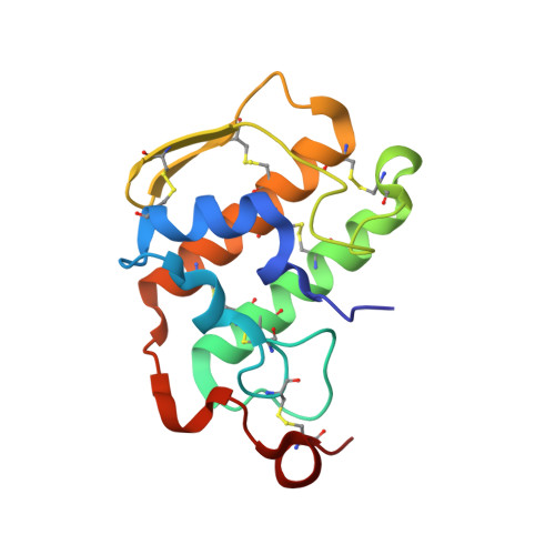
    Pancreatic phospholipase A2 (phospholipase A2 group 1B, G1B) belongs to the superfamily of secreted phospholipase A2 (PLA2) enzymes. G1B has been proposed to be a potential target for diseases such as hypertension, obesity, and diabetes. Human pancreatic prophospholipase A2 (pro-hG1B) is activated by cleavage of the first seven-residue propeptide (phospholipase A2 propeptide, PROP). However, questions still remain on the mode of action for pro-hG1B. In this work, we expressed pro-hG1B in Pichia pastoris and determined the crystal structure at 1.55-A resolution. The x-ray structure demonstrates that pro-hG1B forms a trimer. In addition, PROP occupies the catalytic cavity and can be self-cleaved at 37 degrees C. A new membrane-bound surface and activation mechanism are proposed based on the trimeric model of pro-hG1B. We also propose a new autoproteolytic mechanism for pro-hG1B by the reaction triad Asp49-Arg0-Ser(-2) that is similar to the serine protease catalytic triad.


  • Organizational Affiliation

    From the Guangzhou Institute of Biomedicine and Health, Chinese Academy of Sciences, Guangzhou 510663, China; Graduate University of Chinese Academy of Sciences, Beijing 100049, China.


Macromolecules
Find similar proteins by:  (by identity cutoff)  |  3D Structure
Entity ID: 1
MoleculeChains Sequence LengthOrganismDetailsImage
Phospholipase A2133Homo sapiensMutation(s): 0 
Gene Names: PLA2G1B
EC: 3.1.1.4
Membrane Entity: Yes 
UniProt & NIH Common Fund Data Resources
Find proteins for P04054 (Homo sapiens)
Explore P04054 
Go to UniProtKB:  P04054
PHAROS:  P04054
GTEx:  ENSG00000170890 
Entity Groups  
Sequence Clusters30% Identity50% Identity70% Identity90% Identity95% Identity100% Identity
UniProt GroupP04054
Sequence Annotations
Expand
  • Reference Sequence
Small Molecules
Ligands 1 Unique
IDChains Name / Formula / InChI Key2D Diagram3D Interactions
SO4
Query on SO4

Download Ideal Coordinates CCD File 
B [auth A]SULFATE ION
O4 S
QAOWNCQODCNURD-UHFFFAOYSA-L
Experimental Data & Validation

Experimental Data

  • Method: X-RAY DIFFRACTION
  • Resolution: 1.55 Å
  • R-Value Free: 0.183 
  • R-Value Work: 0.145 
  • R-Value Observed: 0.147 
  • Space Group: P 63
Unit Cell:
Length ( Å )Angle ( ˚ )
a = 56.549α = 90
b = 56.549β = 90
c = 60.631γ = 120
Software Package:
Software NamePurpose
SCALAdata scaling
MOLREPphasing
REFMACrefinement
PDB_EXTRACTdata extraction
CrysalisProdata reduction

Structure Validation

View Full Validation Report


Entry History 

Deposition Data

Revision History  (Full details and data files)

  • Version 1.0: 2009-04-14
    Type: Initial release
  • Version 1.1: 2011-07-13
    Changes: Advisory, Version format compliance
  • Version 1.2: 2014-02-05
    Changes: Database references