3FFB

Gi-alpha-1 mutant in GDP bound form


Experimental Data Snapshot

  • Method: X-RAY DIFFRACTION
  • Resolution: 2.57 Å
  • R-Value Free: 0.226 
  • R-Value Work: 0.181 

wwPDB Validation   3D Report Full Report


Ligand Structure Quality Assessment 


This is version 1.3 of the entry. See complete history


Literature

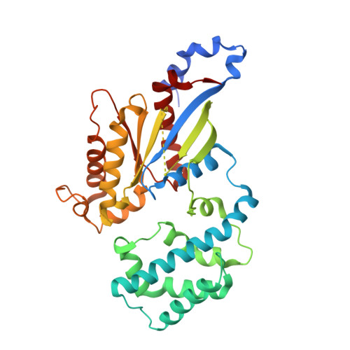
Structural evidence for a sequential release mechanism for activation of heterotrimeric g proteins.

Kapoor, N.Menon, S.T.Chauhan, R.Sachdev, P.Sakmar, T.P.

(2009) J Mol Biol 393: 882-897

  • DOI: https://doi.org/10.1016/j.jmb.2009.08.043
  • Primary Citation of Related Structures:  
    3FFA, 3FFB

  • PubMed Abstract: 

    Heptahelical G-protein (heterotrimeric guanine nucleotide-binding protein)-coupled receptors couple to heterotrimeric G proteins to relay extracellular signals to intracellular signaling networks, but the molecular mechanism underlying guanosine 5'-diphosphate (GDP) release by the G protein alpha-subunit is not well understood. Amino acid substitutions in the conserved alpha5 helix of G(i), which extends from the C-terminal region to the nucleotide-binding pocket, cause dramatic increases in basal (receptor-independent) GDP release rates. For example, mutant Galpha(i1)-T329A shows an 18-fold increase in basal GDP release rate and, when expressed in culture, it causes a significant decrease in forskolin-stimulated cAMP accumulation. The crystal structure of Galpha(i1)-T329A.GDP shows substantial conformational rearrangement of the switch I region and additional striking alterations of side chains lining the catalytic pocket that disrupt the Mg(+2) coordination sphere and dislodge bound Mg(+2). We propose a "sequential release" mechanism whereby a transient conformational change in the alpha5 helix alters switch I to induce GDP release. Interestingly, this mechanistic model for heterotrimeric G protein activation is similar to that suggested for the activation of the plant small G protein Rop4 by RopGEF8.


  • Organizational Affiliation

    Laboratory of Biochemistry and Molecular Biology, Rockefeller University, 1230 York Avenue, New York, NY 10065, USA.


Macromolecules
Find similar proteins by:  (by identity cutoff)  |  3D Structure
Entity ID: 1
MoleculeChains Sequence LengthOrganismDetailsImage
Guanine nucleotide-binding protein G(i), alpha-1 subunit360Rattus norvegicusMutation(s): 1 
Gene Names: Gi-alpha-1Gnai-1Gnai1
UniProt
Find proteins for P10824 (Rattus norvegicus)
Explore P10824 
Go to UniProtKB:  P10824
Entity Groups  
Sequence Clusters30% Identity50% Identity70% Identity90% Identity95% Identity100% Identity
UniProt GroupP10824
Sequence Annotations
Expand
  • Reference Sequence
Small Molecules
Ligands 2 Unique
IDChains Name / Formula / InChI Key2D Diagram3D Interactions
GDP
Query on GDP

Download Ideal Coordinates CCD File 
B [auth A]GUANOSINE-5'-DIPHOSPHATE
C10 H15 N5 O11 P2
QGWNDRXFNXRZMB-UUOKFMHZSA-N
SO4
Query on SO4

Download Ideal Coordinates CCD File 
C [auth A]SULFATE ION
O4 S
QAOWNCQODCNURD-UHFFFAOYSA-L
Experimental Data & Validation

Experimental Data

  • Method: X-RAY DIFFRACTION
  • Resolution: 2.57 Å
  • R-Value Free: 0.226 
  • R-Value Work: 0.181 
  • Space Group: I 4
Unit Cell:
Length ( Å )Angle ( ˚ )
a = 121.761α = 90
b = 121.761β = 90
c = 68.714γ = 90
Software Package:
Software NamePurpose
ADSCdata collection
PHASERphasing
REFMACrefinement
MOSFLMdata reduction
SCALAdata scaling

Structure Validation

View Full Validation Report


Ligand Structure Quality Assessment 


Entry History 

Deposition Data

Revision History  (Full details and data files)

  • Version 1.0: 2009-10-06
    Type: Initial release
  • Version 1.1: 2011-07-13
    Changes: Version format compliance
  • Version 1.2: 2021-10-20
    Changes: Database references, Derived calculations
  • Version 1.3: 2023-09-06
    Changes: Data collection, Refinement description